
Courtesy: Prof Abhijeet Wahegaonkar, Sancheti Hospital, Pune, India
Wrist Arthroscopy, Indications and Techniques
Overview
- Wrist arthroscopy is a minimally invasive technique that allows direct visualization and treatment of disorders affecting the wrist.
- It belongs to the broader field of small-joint arthroscopy, which also includes arthroscopy of the thumb, metacarpophalangeal joints, interphalangeal joints, foot and ankle, and temporomandibular joint.
- Improvements in optical systems, cameras, and miniature instruments have significantly expanded the diagnostic and therapeutic applications of wrist arthroscopy.
- The technique can provide high-resolution visualization of the joint and allows procedures to be performed through small portals with limited disruption of surrounding soft tissues.
Why Wrist Arthroscopy Is Useful
- Wrist arthroscopy can be used for both diagnosis and treatment.
- Direct visualization may help identify pathology that is difficult to evaluate through clinical examination and conventional imaging alone.
- The arthroscopic probe functions much like an extension of the surgeon’s finger, allowing assessment of:
- Cartilage consistency and surface abnormalities.
- Ligament tension and stability.
- Joint congruity.
- Articular surface irregularities.
- Reduction of fracture fragments.
- Articular step-offs.
- The technique has evolved considerably and is now used for a wide range of soft-tissue, ligament, cartilage, and bone conditions.
Equipment and Instrumentation
- Wrist arthroscopy requires smaller and more delicate equipment than many large-joint arthroscopic procedures.
- Commonly used equipment includes:
- A small-diameter arthroscope, commonly 1.9 or 2.9 millimeters in diameter.
- A 30-degree viewing angle, with wider-angle scopes available for selected applications.
- A blunt trocar and cannula to reduce the risk of cartilage injury during entry.
- A small arthroscopic probe.
- Basket and grasping forceps.
- Fine haemostatic or hemostat forceps.
- A small scalpel blade for portal creation.
- Small shavers, burrs, and radiofrequency instruments can be added when more advanced therapeutic procedures are required.
- Modern high-definition optical systems have substantially improved visualization compared with earlier arthroscopic equipment.
Patient Positioning and Joint Distraction
- The patient is generally positioned supine on the operating table.
- Wrist arthroscopy can be performed under regional or general anesthesia, depending on the procedure and clinical circumstances.
- A tourniquet may be used when appropriate.
- The shoulder is commonly abducted to approximately 90 degrees and the elbow flexed to approximately 90 degrees.
- Unlike many arthroscopic procedures in which joint space is created primarily through fluid distension, wrist arthroscopy commonly uses controlled traction or distraction.
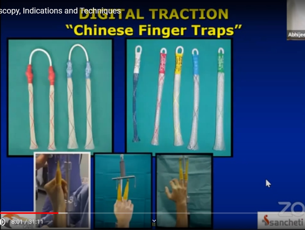
- Finger traps, hand holders, or specialized traction systems can be used to apply distraction.
- Distraction creates sufficient working space within the wrist and facilitates visualization and instrumentation.
- The amount of traction should be carefully controlled to avoid unnecessary soft-tissue or nerve-related complications.
Arthroscopic Portals and Surface Anatomy
- Knowledge of wrist surface anatomy is essential for safe portal placement.
- Most commonly used wrist arthroscopy portals are located dorsally in relation to the extensor tendon compartments.
- Portal names are generally based on their relationship to the extensor compartments.
- Common portals include:
- 3–4 portal.
- 4–5 portal.
- 6R portal.
- 6U portal.
- Radial midcarpal portal.
- Ulnar midcarpal portal.
- The 3–4 portal is positioned between the third and fourth extensor compartments.
- The 4–5 portal lies between the fourth and fifth extensor compartments.
- The 6R portal is positioned immediately radial to the sixth extensor compartment.
- Additional portals can be used according to the pathology and the specific procedure being performed.
Important Anatomical Landmarks
- Careful identification of surface landmarks helps reduce the risk of injury to tendons, nerves, and other soft tissues.
- Lister’s tubercle is an important palpable bony landmark on the dorsal distal radius.
- The distal radius, ulnar styloid, extensor tendons, and metacarpal bases can be identified before portal placement.
- The soft-tissue interval around the wrist can then be used to determine the appropriate portal location.
- After making a small skin incision, blunt spreading rather than deep sharp dissection helps protect underlying structures.
Access to the Wrist Compartments
- Arthroscopy can provide access to several wrist compartments, including:
- Radiocarpal joint.
- Midcarpal joint.
- Distal radioulnar joint.
- Selected carpometacarpal joints.
- The choice of portal depends on the compartment being examined and the pathology being treated.
- Accurate portal placement is particularly important because the wrist contains closely packed tendons, ligaments, nerves, and vascular structures.
Diagnostic Arthroscopy
- A systematic examination helps minimize the possibility of overlooking pathology.
- A commonly used sequence is:
- Examination of the radiocarpal joint.
- Examination from the radial side toward the ulnar side.
- Assessment from distal to proximal.
- Examination of palmar and dorsal structures.
- Evaluation of ligaments and cartilage.
- Although there is no single mandatory sequence, following a consistent examination pattern improves the reliability of diagnostic arthroscopy.
Radiocarpal Joint
Important structures that may be evaluated include:
- Distal radius.
- Scaphoid fossa.
- Lunate fossa.
- Scapholunate ligament.
- Triangular fibrocartilage complex.
- Radiocarpal ligaments.
- Cartilage surfaces.
- Synovial tissue.
- Dorsal and palmar portions of relevant ligamentous structures.
Midcarpal Joint
The midcarpal examination allows assessment of:
• Scaphoid.
• Lunate.
• Triquetrum.
• Capitate.
• Hamate.
• Scapholunate ligament.
• Lunotriquetral ligament.
• Other intrinsic and extrinsic ligamentous structures.
• Carpometacarpal joint relationships.
Major Therapeutic Applications
The therapeutic role of wrist arthroscopy has expanded substantially. Important applications include:
- Triangular fibrocartilage complex injuries.
- Intrinsic ligament injuries.
- Distal radius fractures involving the joint surface.
- Scaphoid fractures.
- Scaphoid non-union.
- Ganglion cyst excision.
- Ulnocarpal impaction syndrome.
- Selected procedures for wrist arthritis.
- Synovectomy and debridement.
- Selected reconstructive and stabilizing procedures.
Triangular Fibrocartilage Complex Injuries
- Arthroscopy allows direct assessment of the triangular fibrocartilage complex and helps determine the location and characteristics of a tear.
- A probe can be used to evaluate the stability of the tissue and identify peripheral tears.
- Arthroscopic treatment may include debridement or repair depending on the tear pattern, tissue quality, and associated instability.
- Peripheral tears may be treated with arthroscopic capsular or ligamentous repair techniques.
- Tears involving the foveal attachment may require reinsertion of the triangular fibrocartilage complex to its anatomical attachment using appropriate fixation methods.
- Successful repair aims to restore the stability of the distal radioulnar joint when instability is present.
Scapholunate Ligament Injuries
- Arthroscopy provides direct assessment of the scapholunate ligament from both the radiocarpal and midcarpal compartments.
- The extent of ligament injury and the condition of the remaining tissue can be evaluated using a probe.
- Selected ligament tears can be treated with arthroscopic repair techniques.
- Depending on the injury pattern, sutures may be passed through the remaining ligamentous tissue and capsule to restore the relationship between the scaphoid and lunate.
- Temporary fixation may sometimes be used to protect the repair while healing occurs.
- Treatment is individualized according to the stage and severity of instability.
Distal Radius Fractures
- Arthroscopy can assist in the management of fractures extending into the wrist joint.
- It is particularly useful when accurate restoration of the articular surface is important.
- Arthroscopic visualization can help:
- Assess the joint surface.
- Improve fracture reduction.
- Identify residual articular incongruity.
- Detect associated ligament or triangular fibrocartilage complex injuries.
- Confirm implant position.
- Arthroscopy may therefore complement minimally invasive fracture fixation rather than replace conventional fixation techniques.
Scaphoid Fractures
- Selected scaphoid fractures can be reduced and stabilized using minimally invasive techniques assisted by arthroscopic visualization.
- Arthroscopy allows direct assessment of fracture reduction and fixation.
- It can help identify whether a screw is appropriately positioned and whether any portion of the implant projects into the joint.
- Preserving the surrounding soft-tissue envelope may be beneficial when performing minimally invasive fixation.
Scaphoid Non-union
- Arthroscopic techniques can be used in selected cases of scaphoid non-union.
- Bone grafting may be performed through a small portal under arthroscopic visualization.
- Cancellous bone graft can be introduced into the non-union site and carefully packed into the defect.
- Care is required to prevent graft material from entering the joint.
- After grafting, the scaphoid can be stabilized using appropriate percutaneous fixation when indicated.
- Patient selection is important because not all patterns of scaphoid non-union are suitable for arthroscopic treatment.
Ulnocarpal Impaction Syndrome
- Arthroscopic distal ulna resection can be considered in selected patients with ulnocarpal impaction.
- The objective is to reduce excessive contact between the ulna and the carpus.
- This procedure provides an arthroscopic alternative to certain forms of ulnar shortening surgery in appropriately selected cases.
- Preservation of the sigmoid notch and the stability of the distal radioulnar joint are important considerations.
- Excessive bone removal can compromise joint stability and should therefore be avoided.
Ganglion Cyst Excision
- Arthroscopic excision is an established minimally invasive treatment option for selected wrist ganglion cysts.
- Dorsal wrist ganglion excision is also commonly used as an introductory therapeutic procedure during wrist arthroscopy training.
- Arthroscopic treatment allows visualization of the cyst’s relationship to the joint and surrounding structures.
Wrist Arthritis and Carpometacarpal Arthroscopy
- Arthroscopy can also be performed in selected carpometacarpal joint disorders.
- A small-diameter arthroscope can be used to inspect the joint surface and surrounding structures.
- Arthroscopic assessment allows evaluation of:
- Articular cartilage.
- Joint surfaces.
- Ligamentous structures.
- Degenerative changes.
- Selected procedures for carpometacarpal arthritis may include arthroscopic debridement or resection-based procedures, depending on the joint involved and the severity of disease.
Advantages of Wrist Arthroscopy
- Minimally invasive access.
- High-resolution visualization of intra-articular structures.
- Ability to evaluate cartilage and ligament integrity directly.
- Assistance with accurate fracture reduction.
- Ability to diagnose and treat selected conditions during the same procedure.
- Potentially less disruption of surrounding soft tissues than open approaches.
- Ability to identify associated injuries that may not be apparent through external examination alone.
Limitations and Safety Considerations
- Wrist arthroscopy requires detailed knowledge of wrist anatomy and extensive technical training.
- The small working space leaves little margin for error.
- Important structures such as extensor tendons, sensory nerves, cartilage, and ligaments must be protected.
- Portal placement should be based on clearly identified anatomical landmarks.
- Sharp instruments should not be advanced deeply without appropriate control.
- Traction should be carefully regulated.
- Arthroscopy should be considered a specialized technique rather than a substitute for careful clinical assessment and appropriate imaging.
Future Development
- Wrist arthroscopy continues to evolve with advances in imaging, instrumentation, and minimally invasive techniques.
- Smaller and more flexible scopes may improve access to difficult anatomical regions.
- Improved shavers and other miniature instruments may expand the range of therapeutic procedures.
- Continued anatomical and clinical research may identify additional applications for arthroscopic treatment.
- The overall direction of development is toward increasingly precise, minimally invasive procedures that combine detailed visualization with tissue preservation.
Key Takeaways
- Wrist arthroscopy provides both diagnostic and therapeutic access to the small and complex structures of the wrist.
- Successful arthroscopy depends heavily on an accurate understanding of surface and portal anatomy.
- A systematic examination of the radiocarpal and midcarpal joints helps reduce the risk of missing pathology.
- Common applications include treatment of triangular fibrocartilage complex injuries, ligament injuries, selected distal radius and scaphoid fractures, scaphoid non-union, ganglion cysts, and ulnocarpal impaction.
- Arthroscopy can also assist with fracture reduction and assessment of associated ligamentous injuries.
- Modern high-definition optics and miniature instruments have significantly expanded the capabilities of wrist arthroscopy.
- Because of the close proximity of important tendons, nerves, ligaments, and cartilage, the procedure requires specialized training and careful attention to anatomy.
Leave a Reply