
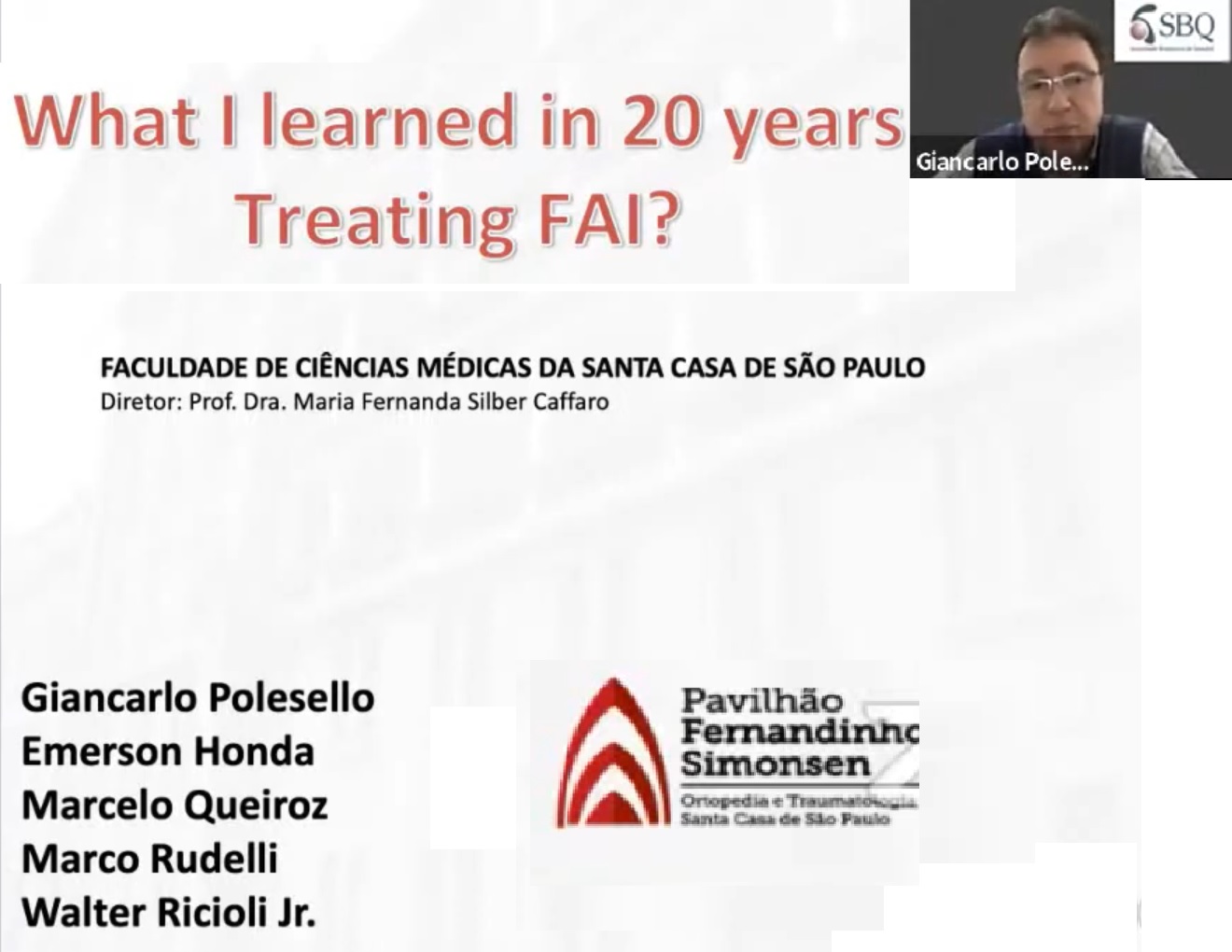
Courtesy: Prof Giancarlo Polesello, MD, PhD,Sao Paolo, Brazil
President, Brazilian Hip Society
Hip Arthroscopy: Practical Lessons in Femoroacetabular Impingement
Overview
- Hip arthroscopy has evolved substantially and is now an established treatment option for appropriately selected patients with symptomatic intra-articular hip disorders.
- The procedure is technically demanding because the hip is a deep joint with:
- A relatively constrained joint space.
- A thick capsule.
- Close proximity to important neurovascular structures.
- Complex three-dimensional anatomy.
- A requirement for specialized instruments, positioning and traction.
- Early experience with hip arthroscopy was limited by inadequate imaging, poorly defined indications, technical difficulties and uncertainty regarding outcomes.
- Increasing experience, improved imaging and better understanding of hip pathology have substantially improved patient selection and surgical techniques.
Learning Curve
- Hip arthroscopy has a significant learning curve.
- The learning process can be considered in 4 stages:
- Unconscious incompetence: The learner is unaware of what is not yet understood.
- Conscious incompetence: The learner recognizes the limitations in knowledge or technical ability.
- Conscious competence: The procedure can be performed successfully but requires considerable concentration.
- Unconscious competence: Technical skills become more automatic through repeated experience.
- Progression through these stages requires:
- Structured training.
- Repeated exposure to appropriate cases.
- Recognition and analysis of complications.
- Supervision and mentorship.
- Persistence during the early learning phase.
- Published data involving 8,041 hip arthroscopies performed by 251 surgeons demonstrated a clear relationship between surgeon experience and the risk of subsequent surgery. Cases performed by surgeons with the highest career volumes had substantially lower rates of additional surgery than those performed by lower-volume surgeons.
- Hip arthroscopy should therefore ideally be developed within an appropriate training environment rather than through unsupervised acquisition of complex skills.
Traction and Joint Access
- Adequate distraction of the hip is essential for safe access to the central compartment.
- The objective of traction is to distract the femoral head from the acetabulum sufficiently to create working space.
- The direction and vector of traction are important.
- Both central-post and eccentric-post traction systems may be used depending on the operating table and surgeon preference.
- Appropriate positioning and traction technique are important to reduce:
- Neurological injury.
- Perineal or pudendal pressure-related complications.
- Excessive traction-related morbidity.
- Traction time should be minimized whenever possible.
Portal Placement
- Commonly used hip arthroscopy portals include:
- Anterolateral portal.
- Anterior portal.
- Posterolateral or posterior portal.
- The anterolateral portal is commonly established initially because of its relative distance from major neurovascular structures.
- Additional portals can subsequently be established under direct visualization.
- Particular attention should be paid to the lateral femoral cutaneous nerve during anterior portal placement.
- Small adjustments in portal position may help reduce the risk of injury to the nerve.
- A portal established initially with limited visualization should subsequently be inspected arthroscopically to identify inadvertent:
- Labral penetration.
- Chondral injury.
- Capsular injury.
- Inappropriate intra-articular positioning.
- Guide needles should be introduced through safe anatomical intervals and directed carefully to avoid the labrum and articular cartilage.
Arthroscopic Anatomy
Important structures encountered during hip arthroscopy include:
- Femoral head.
- Femoral neck.
- Acetabular rim.
- Acetabular articular cartilage.
- Acetabular labrum.
- Ligamentum teres.
- Acetabular fossa.
- Pulvinar tissue.
- Synovium and synovial folds.
- Zona orbicularis.
- Chondrolabral junction.
Important technical principle
- The labrum and articular cartilage should be protected during portal establishment and instrument manipulation.
- Careful identification of normal anatomy is essential before beginning corrective procedures.
Femoroacetabular Impingement
- Femoroacetabular impingement occurs when abnormal contact develops between the proximal femur and acetabulum during hip movement.
- The major morphological patterns are:
- Cam morphology: Abnormal prominence or reduced femoral head-neck offset, particularly affecting the anterosuperior femoral head-neck junction.
- Pincer morphology: Excessive acetabular coverage or other acetabular morphology resulting in abnormal contact.
- Combined morphology: Features of both cam and pincer morphology.
- Repetitive abnormal contact may contribute to:
- Labral injury.
- Chondral injury.
- Chondrolabral separation.
- Cartilage delamination.
- Progressive degenerative changes.
Imaging Must Be Interpreted in Clinical Context
- The presence of cam or pincer morphology on imaging does not by itself establish symptomatic femoroacetabular impingement.
- Morphological findings are common in asymptomatic individuals.
- A systematic review reported cam morphology in approximately 37% and pincer morphology in approximately 67% of asymptomatic hips; labral abnormalities were also frequently identified on magnetic resonance imaging.
- Therefore:
- Imaging findings should not be treated in isolation.
- Symptoms must be correlated with physical examination.
- Alternative causes of pain must be considered.
- A useful clinical principle is:
Treat the patient, not the imaging finding.
Differential Diagnosis of Hip and Groin Pain
Persistent symptoms attributed to femoroacetabular impingement may arise from other conditions.
Important alternatives include:
- Lumbar spine pathology.
- Lumbar disc disease.
- Sacroiliac joint pathology.
- Pubic symphysis disorders.
- Adductor-related groin pain.
- Rectus abdominis–adductor pathology.
- Iliopsoas pathology.
- Greater trochanteric pain syndrome.
- Other extra-articular causes of hip and groin pain.
- In athletes, associated injuries elsewhere in the kinetic chain should also be assessed.
Clinical lesson
- A patient with persistent pain after technically satisfactory hip surgery should undergo a complete reassessment rather than automatically being considered a case of recurrent femoroacetabular impingement.
- Disproportionate pain is particularly important because it may indicate:
- An alternative diagnosis.
- A postoperative complication.
- Neuropathic pain.
- Extra-articular pathology.
- A significant psychological or functional component.
Cartilage Damage and Delamination
- Repetitive femoroacetabular conflict can injure the labrum and adjacent cartilage.
- Chondrolabral disruption may allow synovial fluid to extend beneath partially separated cartilage.
- Progressive cartilage delamination may eventually involve weight-bearing areas.
- Advanced cartilage damage is associated with poorer prognosis.
- Recognition and appropriate treatment of associated cartilage and labral pathology are therefore important components of hip preservation surgery.
Indications for Surgical Treatment
- Surgical treatment should be considered only in appropriately selected symptomatic patients.
- Important considerations include:
- Consistent clinical symptoms.
- Compatible physical examination findings.
- Appropriate imaging abnormalities.
- Failure of appropriate nonoperative management when indicated.
- Absence of advanced degenerative joint disease that would make hip preservation unlikely to succeed.
- Surgical objectives may include:
- Correction of cam morphology.
- Correction of symptomatic acetabular overcoverage where appropriate.
- Restoration or preservation of the labrum.
- Treatment of focal cartilage lesions.
- Removal of mechanical sources of impingement.
- Hip arthroscopy can improve pain and function in appropriately selected patients.
- It should not be represented as a guaranteed method of preventing future osteoarthritis or total hip arthroplasty.
Causes of Failed Hip Arthroscopy
Failure after hip arthroscopy may result from multiple factors.
Patient-selection factors
- Incorrect diagnosis.
- Symptoms arising from extra-articular pathology.
- Advanced cartilage damage.
- Established osteoarthritis.
- Inappropriate expectations.
Technical factors
- Residual cam morphology.
- Residual pincer morphology.
- Inadequate osteoplasty.
- Over-resection.
- Unrecognized labral pathology.
- Unrecognized or inadequately treated cartilage damage.
- Intra-articular implant or anchor complications.
Biological factors
- Failure of labral healing.
- Progression

Leave a Reply