Courtesy: Amr Abdelgawad, Maimonaides Medical Centre, Brooklyn, New York
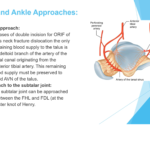
Posterior Approach to the Glenoid
- Used for fixation of posterior glenoid fractures and access to the posterior capsule.
- Internervous plane: between infraspinatus (suprascapular nerve) and teres minor (axillary nerve).
- This interval provides safe access to the posterior glenoid.
- Axillary nerve runs inferior to teres minor and must be protected.
- Excessive superior retraction of infraspinatus may stretch the suprascapular nerve and artery.
- Infraspinatus splitting approach can be used for direct access to the posterior capsule with lower risk to the axillary nerve.
Deltopectoral Approach
- Commonly used approach for proximal humerus fractures and shoulder arthroplasty.
- Interval: between deltoid muscle (laterally) and pectoralis major muscle (medially).
- Subscapularis tendon must be divided or lesser tuberosity osteotomy performed to enter the joint.
- Repair of subscapularis is required at the end of surgery.
- Postoperative precautions: limit active internal rotation and excessive passive external rotation to protect repair.
Lateral Deltoid Splitting Approach
- Used for fixation of proximal humerus fractures.
- Approach splits the deltoid muscle fibers.
- Axillary nerve runs approximately 5 cm distal to the lateral edge of the acromion.
- Incision must be limited or the axillary nerve identified before extending distally.
Posterior Approach to the Humerus
- Used for open reduction and internal fixation of humeral shaft fractures.
- Traditional approach: triceps splitting technique after identification of the radial nerve.
- Modified approach: triceps-sparing technique where medial and lateral heads are mobilized.
- Traditional approach exposes approximately 60–65% of the humeral shaft.
- Modified approach exposes up to 95% of the humeral shaft.
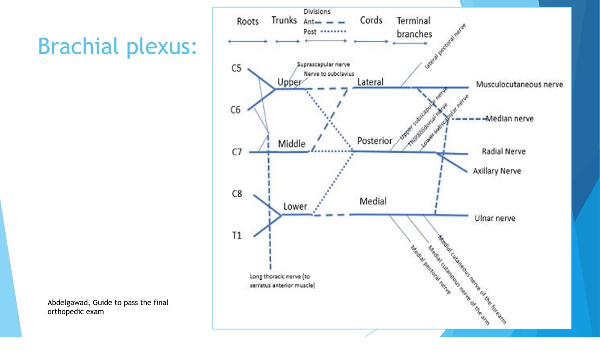
Brachial Plexus Overview
- Brachial plexus provides motor and sensory innervation to the upper limb.
- Formed from nerve roots C5, C6, C7, C8, and T1.
- Organization follows: Roots ? Trunks ? Divisions ? Cords ? Terminal branches.
Trunks of the Brachial Plexus
- C5–C6 form the upper trunk.
- C7 continues as the middle trunk.
- C8–T1 form the lower trunk.
Divisions
- Each trunk divides into anterior and posterior divisions.
- Anterior divisions generally supply flexor compartments.
- Posterior divisions generally supply extensor compartments.
Cords of the Brachial Plexus
- Posterior cord: formed by posterior divisions of all three trunks.
- Lateral cord: formed by anterior divisions of upper and middle trunks.
- Medial cord: formed by anterior division of the lower trunk.
Major Branches
- Long thoracic nerve (C5–C7): supplies serratus anterior.
- Upper trunk branches: suprascapular nerve and nerve to subclavius.
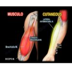
- Lateral cord: lateral pectoral nerve, musculocutaneous nerve, and lateral contribution to median nerve.
- Posterior cord: axillary nerve, radial nerve, thoracodorsal nerve, upper and lower subscapular nerves.
- Medial cord: ulnar nerve, medial pectoral nerve, medial cutaneous nerves of arm and forearm, medial contribution to median nerve.
Important Shoulder Nerve Supply
- Axillary nerve supplies deltoid and teres minor.
- Suprascapular nerve supplies supraspinatus and infraspinatus.
- Upper and lower subscapular nerves supply subscapularis.
- Lower subscapular nerve also supplies teres major.
Spaces Around the Shoulder
- Quadrangular space: bounded by teres minor (superior), teres major (inferior), long head of triceps (medial), and humerus (lateral).
- Contents: axillary nerve and posterior circumflex humeral artery.
- Triangular space: bounded by teres minor, teres major, and long head of triceps.
- Contents: circumflex scapular artery.
- Triangular interval: bounded by teres major, long head of triceps, and lateral head of triceps.
- Contents: radial nerve and profunda brachii artery.
Rotator Interval
- Triangular space in the anterior-superior shoulder joint capsule.
- Boundaries: supraspinatus (superior), subscapularis (inferior), coracoid process (base), transverse humeral ligament (apex).
- Contents: long head of biceps tendon, coracohumeral ligament, and superior glenohumeral ligament.
- Important for stability of the glenohumeral joint.
Glenohumeral Ligament Functions
- Coracohumeral ligament: primary restraint to inferior translation of the humeral head.
- Superior glenohumeral ligament: limits inferior translation with arm adducted.
- Middle glenohumeral ligament: limits anterior translation in mid-abduction (~45°).
- Inferior glenohumeral ligament complex: primary stabilizer at 90° abduction.
Blood Supply of the Humeral Head
- Posterior circumflex humeral artery provides approximately 60% of blood supply.
- Anterior circumflex humeral artery contributes approximately 40%.
- Ascending branch of the anterior circumflex artery forms the arcuate artery.
Rotator Cuff Footprint
- Supraspinatus insertion footprint approximately 16 mm mediolateral.
- Exposure of ~7–8 mm of bone during arthroscopy suggests about 50% tendon involvement.
Biceps Tendon and Bicipital Groove
- Long head of biceps tendon runs through the bicipital groove.
- Lateral lip: insertion of pectoralis major.
- Medial lip: insertion of teres major.
- Floor of groove: insertion of latissimus dorsi.
- Biceps pulley formed by coracohumeral ligament and superior glenohumeral ligament prevents medial subluxation.

Leave a Reply