
Courtesy: Prof Nabil Ebraheim, University of Toledo, Ohio, USA
Clinical Examination of Low Back Pain
Diagnostic Scope & Anatomical Generators
Low back pain requires systematic clinical evaluation to distinguish among anatomical pain generators:
-
Intervertebral discs
-
Lumbar nerve roots
-
Sacroiliac (SI) joints
-
Facet joints
-
Paraspinal musculature and ligaments
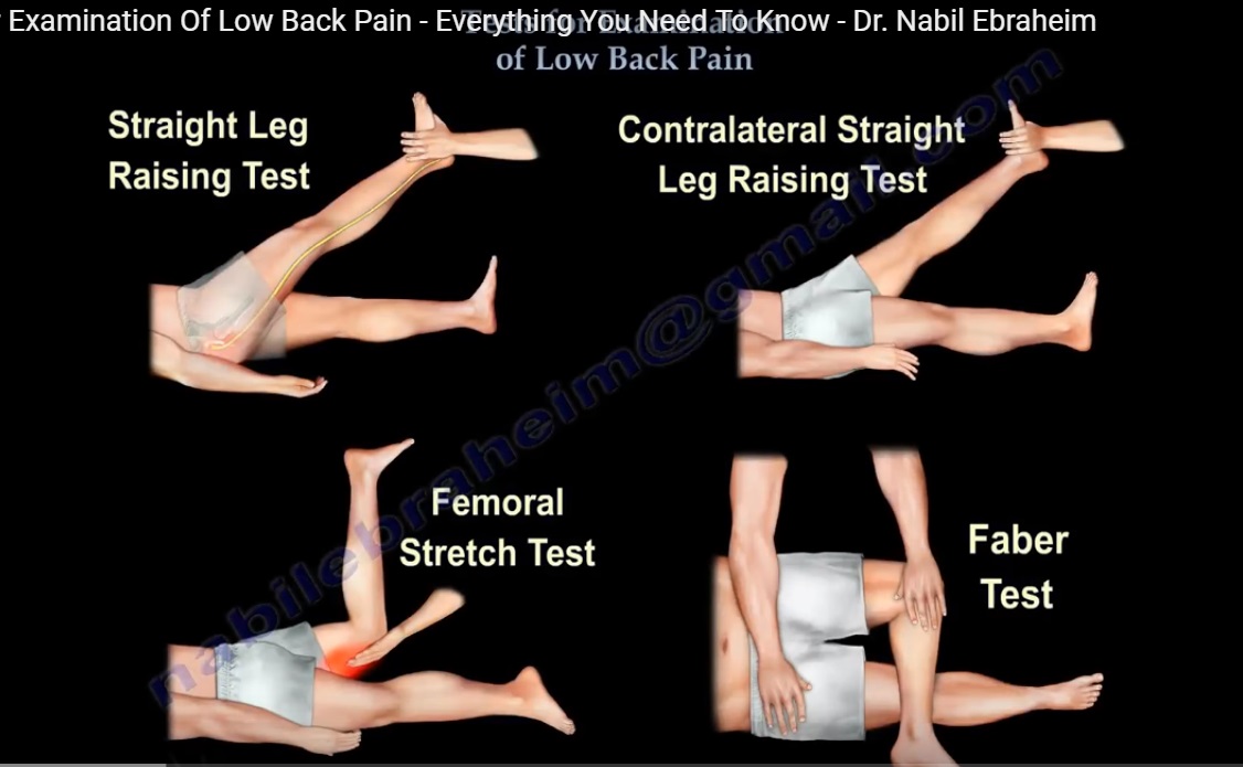
1. Straight Leg Raising (SLR) Test
-
Clinical Indication: Provocation of sciatic and lower lumbar nerve-root irritation (predominantly L5, S1).
-
Technique:
-
Patient is placed supine.
-
Symptomatic leg is elevated with the knee maintained in full extension.
-
Hip flexion progressively tensions the sciatic nerve and its arborizing roots.
-
-
Positive Criteria: Reproduction of true radicular pain radiating distal to the knee at $<60^\circ$ of hip flexion. (Isolated low-back pain is considered a negative test).
-
Neural Tension Modifications:
-
Knee Flexion: Flexing the knee abolishes sciatic tension and relieves radicular pain, corroborating a neurogenic pain etiology.
-
Ankle Dorsiflexion (Bragard Maneuver): Lowering the leg slightly below the painful threshold and adding ankle dorsiflexion re-tensions the sciatic nerve, reproducing radicular symptoms.
-
2. Contralateral (Crossed) Straight Leg Raising Test
-
Technique: Passive elevation of the asymptomatic (unaffected) lower extremity with the knee in full extension.
-
Positive Criteria: Reproduction of back and radiating radicular pain in the contralateral, symptomatic limb.
-
Clinical Significance: Highly indicative of a large, extruded, or sequestered disc herniation.
3. Femoral Nerve Stretch Test (Reverse SLR)
-
Clinical Indication: Assessment of upper lumbar radiculopathy (primarily L3 and L4 roots) and femoral nerve irritation.
-
Technique: Patient positioned prone or lateral; the hip is passively extended with the knee flexed to tension the femoral nerve.
-
Positive Criteria: Reproduction of sharp pain along the ipsilateral anterior thigh.
-
Clinical Utility: Less commonly positive/utilized than standard SLR because lumbar disc herniations overwhelmingly involve L5 and S1.
4. Sacroiliac Joint Evaluation: FABER Test & Diagnostic Blocks
-
FABER (Flexion–ABduction–External Rotation) Maneuver:
-
Technique: Supine patient; the involved hip is positioned in flexion, abduction, and external rotation with the foot resting over the contralateral knee. Downward pressure is applied concurrently to the flexed ipsilateral knee and the contralateral anterior superior iliac spine (ASIS).
-
Positive Criteria: Provocation of pain localized to the posterior sacroiliac region.
-
Diagnostic Limitation: Suggestive but non-confirmatory. Maneuver stresses overlapping pain generators including the lumbar disc, facets, neural elements, muscles, and surrounding ligaments.
-
-
Diagnostic SI Joint Infiltration:
-
Diagnostic injection using local anesthetic with or without corticosteroid.
-
Transient symptomatic pain relief supports the SI joint as the primary symptomatic generator.
-
-
Clinical Algorithm: Recommended when localized unilateral low-back pain is accompanied by a positive FABER test and high clinical suspicion.
5. Directional Lumbar Motion Testing
-
Lumbar Flexion:
-
Biomechanical Effect: Increases neuroforaminal and spinal canal dimensions.
-
Clinical Correlation: Pain provocation indicates discogenic pathology.
-
-
Lumbar Extension:
-
Biomechanical Effect: Decreases neuroforaminal and canal cross-sectional area; loads posterior elements.
-
Clinical Correlation: Pain provocation suggests lumbar canal stenosis, facet arthropathy, spondylolysis, or spondylolisthesis.
-
-
Shopping Cart Phenomenon: Relief of neurogenic claudication/stenotic symptoms achieved by leaning forward or flexing the trunk, attributable to expansion of the neuroforaminal space.
6. Upper Motor Neuron (UMN) / Myelopathy Signs
-
Clinical Rationale: Lower lumbar disc lesions cause lower motor neuron (LMN) root compression. The presence of UMN signs mandates evaluation of the cervical and thoracic cord, irrespective of concomitant lumbar MRI findings.
-
Pathological Signs:
-
Ankle Clonus: Elicited by abrupt, sustained passive ankle dorsiflexion. Sustained rhythmic oscillations denote UMN disinhibition.
-
Babinski Reflex: Stroke along the lateral plantar foot margin up to the base of the metatarsals. Pathological response is great toe extension accompanied by fanning of the lesser digits.
-
Gait Patterns: Myelopathic or spastic gait disturbances.
-
7. Evaluation of Non-Organic Pathology (Waddell Signs)
Waddell signs assist in identifying behavioral or non-organic components in chronic low back pain:
-
Simulation Testing: Axial loading of the skull or passive trunk rotation that produces low-back pain despite minimal mechanical lumbar strain.
-
Tenderness: Superficial or non-anatomical/widespread tenderness.
-
Distraction Testing (e.g., Flip Test): Inconsistency between formal supine SLR and seated straight leg extension.
-
Regional Disturbances: Non-dermatomal sensory loss or generalized, non-myotomal muscle weakness (“cogwheeling”).
-
Overreaction: Disproportionate behavioral, vocal, or physical display during examination.
-
Interpretation: Non-organic signs identify altered pain behavior, potential exaggeration, or secondary gain; they do not substantiate malingering or prove the total absence of organic pathology.
Master Summary & Core Clinical Correlates
-
Straight Leg Raising Test: L5/S1 root tension; radicular leg pain elicited at $<60^\circ$.
-
Crossed Straight Leg Raising Test: Suggests large, extruded, or sequestered lumbar disc herniation.
-
Femoral Nerve Stretch Test: Evaluates upper lumbar roots (L3, L4); positive when reproducing anterior thigh pain.
-
FABER Test: Suggestive screening maneuver for SI joint pathology; confirmed by diagnostic local block.
-
Flexion-Aggravated Pain: Discogenic etiology.
-
Extension-Aggravated Pain: Facet joint arthrosis, lumbar canal stenosis, spondylolysis, or spondylolisthesis.
-
Shopping Cart Sign: Flexion-mediated relief in lumbar canal stenosis due to increased foraminal volume.
-
Myelopathy Screening (Babinski, Clonus): Indicates spinal cord (cervical/thoracic) pathology; not explained by standard lumbar radiculopathy.
-
Waddell Signs: Identify non-organic or behavioral components; controversial and distinct from deliberate malingering.
Leave a Reply