
Courtesy: Prof Nabil Ebraheim, University of Toledo, Ohio, USA
Meniscal Tear — Clinical Examination Tests
Overview
The meniscus acts as an important cushion between the femoral condyle and tibial plateau.
A typical meniscal injury history includes:
- Twisting injury
- Medial or lateral joint-line pain
- Swelling/effusion
- Mechanical symptoms:
- Clicking
- Locking
- Catching
Range of motion is usually normal, except in some mechanically blocked tears.
- Most Important Clinical Finding
The most important examination finding emphasized in is:
Joint-line tenderness
Tenderness may be along the:
- Medial joint line
- Lateral joint line
and should correspond to the symptomatic side.
However, joint-line pain can occasionally arise from other causes such as:
- Bursitis
- Arthritis
- Ligament injury
So it is suggestive, not absolutely diagnostic.
- Principle of Provocative Meniscal Tests
The different meniscal tests use the same basic principle:
Trap the meniscus between the femur and tibia while applying rotation/twisting.
The tests differ mainly by patient position:
- Supine
- Prone
- Standing
A provocative test is suggestive of meniscal tear when it produces:
Pain with a click or pop
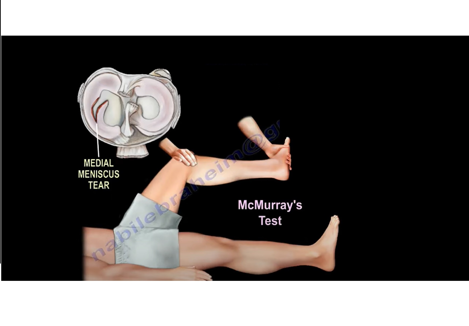
- McMurray Test
identifies the McMurray test as the most popular provocative test for meniscal pathology.
Its principle is:
Knee movement + tibiofemoral rotation – compression/trapping of meniscus – pain/click if torn.
The detailed medial-versus-lateral maneuver is not provided in the supplied transcript, so it should not be added here from outside knowledge.
- Apley Compression Test
“ably compression test” refers to the Apley compression test.
Position
- Patient lies prone
- Knee flexed to 90°
Technique
- Apply axial compression
- Rotate the tibia internally and externally
- Palpate/monitor the joint line
Positive test
Joint-line pain with a click or pop
suggests meniscal pathology.
Why is it called a compression test?
Because the tibia is compressed toward the femur, loading the meniscus.
- Apley Compression vs Distraction
This distinction is very important.
| Maneuver | Interpretation in transcript |
| Rotation + compression | Meniscal injury |
| Rotation + distraction | Ligamentous injury |
Thus:
Pain with compression – think meniscus
Pain with distraction – think ligament
- Thessaly Test
The final standing test described is the Thessaly test.
Position
- Patient stands on the affected leg
- Knee flexed approximately 20°
- Examiner holds the patient’s hands for support
Technique
The patient rotates/twists the knee:
- Internal rotation
- External rotation
about three times.
Positive test
Pain with clicking or popping
suggests a meniscal tear.
- Comparison of Meniscal Provocative Tests
| Test | Patient position | Basic maneuver | Positive finding |
| McMurray | Supine | Knee motion + rotation | Pain/click/pop |
| Apley compression | Prone | Compression + tibial rotation | Pain/click/pop |
| Thessaly | Standing | Weight-bearing rotation at ~20° flexion | Pain/click/pop |
- Why These Tests Work
All three try to reproduce the same mechanical event:
Rotation/twisting – meniscus trapped between femoral condyle and tibial plateau – torn segment becomes painful or produces click/pop.
The main difference is the way the knee is loaded.
- Mechanical Symptoms
Symptoms supporting a meniscal tear include:
- Clicking
- Catching
- Locking
These are especially important when associated with:
- Twisting injury
- Joint-line pain
- Joint-line tenderness
- Bucket-Handle Tear
A bucket-handle tear can mechanically block the knee.
Important clinical clue:
Loss of full extension / locked knee
The speaker recommends obtaining MRI early when a bucket-handle tear is suspected because of the mechanical block.
- Role of MRI
emphasizes that provocative meniscal tests are not fully specific.
Therefore:
MRI is used to confirm a suspected meniscal tear.
MRI becomes particularly important when there are:
- Mechanical symptoms
- Persistent symptoms
- Suspected bucket-handle tear
- Loss of extension
High-Yield Diagnostic Pattern
Typical meniscal tear
Twisting injury
Medial/lateral joint-line pain
Effusion ± clicking/catching/locking
Joint-line tenderness
Positive provocative test
MRI confirmation
Key Take-Home Points
- Joint-line tenderness is the most important clinical finding emphasized in the transcript.
- Meniscal provocative tests all attempt to trap the meniscus between femur and tibia with rotation.
- McMurray is described as the most popular test.
- Apley compression: compression + rotation – meniscal pain.
- Apley distraction: pain suggests ligamentous pathology rather than meniscal pathology.
- Thessaly: standing on affected limb with knee around 20° flexion, then twisting.
- A positive provocative test typically produces pain with click/pop.
- These tests are not completely specific.
- MRI is used to confirm meniscal tear.
- Bucket-handle tear may block full extension, prompting early MRI.
Exam Pearls
Meniscus – joint-line tenderness
McMurray – supine
Apley – prone
Thessaly – standing
Compression hurts – meniscus
Distraction hurts – ligament
Locked knee + loss of extension – think bucket-handle tear
Leave a Reply