

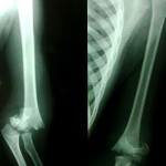
Courtesy: Prof Nabil Ebraheim,University of Toledo, Ohio, USA
Supracondylar Fracture of the Humerus: Circulatory Assessment and Management
Initial Principles
- Perform a thorough neurovascular examination before and after reduction.
- Avoid elbow hyperflexion beyond 90 degrees during casting because it may compress the brachial artery.
- Closed reduction and percutaneous pinning (CRPP) is the preferred treatment.
- The most feared vascular complication is Volkmann ischemic contracture secondary to brachial artery injury.
Vascular Assessment
Pulseless but Well Perfused (Pink) Hand
Clinical features
- Absent radial pulse.
- Warm, pink hand.
- Normal capillary refill.
Management
- Perform urgent closed reduction and percutaneous pinning.
- Observe for 24 to 48 hours.
- Repeat serial neurovascular examinations.
Rationale
- Adequate collateral circulation maintains hand perfusion despite absence of the radial pulse.
Pulseless and Poorly Perfused (Cold) Hand
Clinical features
- Absent radial pulse.
- Cold hand.
- Pale or cyanotic appearance.
- Delayed capillary refill.
Management
- Proceed immediately to the operating room.
- Perform closed reduction and percutaneous pinning.
- Reassess circulation after reduction.
- If perfusion does not improve, perform immediate brachial artery exploration.
Key point
- Fracture reduction should always be performed before vascular exploration.
- Management principles are similar to vascular injuries associated with knee dislocation.
Loss of Perfusion During Reduction or Observation
Clinical features
- Initially well perfused hand becomes ischemic.
Management
- Perform immediate open exploration of the brachial artery.
- Do not delay treatment with arteriography.
Loss of Circulation After Pinning
Possible causes
- Vascular compromise caused by fracture reduction.
- Entrapment of the neurovascular bundle by fracture fragments or pins.
Management
- Remove the pins.
- Reverse the reduction.
- Reassess circulation.
- If perfusion remains compromised, perform brachial artery exploration.
Intraoperative Surgical Pearls
- Inspect the fracture site for a persistent fracture gap during reduction.
- A persistent gap may indicate interposition or entrapment of the neurovascular bundle.
Management Algorithm
Pulseless but well perfused hand
- Closed reduction and percutaneous pinning.
- Observe with serial neurovascular assessment.
Pulseless and poorly perfused hand
- Closed reduction and percutaneous pinning.
- Reassess perfusion.
- If circulation does not improve, proceed to immediate brachial artery exploration.
Perfusion lost during reduction or observation
- Immediate vascular exploration.
Perfusion lost after pinning
- Remove pins.
- Reverse the reduction.
- Reassess circulation.
- Explore the brachial artery if vascular compromise persists.
Viva Pearls
- A pulseless pink hand should be treated with fracture fixation followed by close observation.
- A cold pulseless hand is a surgical emergency.
- Fracture reduction is the first step in the management of vascular compromise.
- Avoid hyperflexion casting because it can compress the brachial artery.
- The brachial artery is the vessel most commonly injured in supracondylar fractures of the humerus.


Best education