1. Common Pediatric Elbow Injuries
- Supracondylar fracture of humerus (most common)
- Lateral condyle fracture of humerus
- Medial epicondyle fracture
- Elbow dislocation
- Radial neck fracture
- Olecranon fracture
2. Supracondylar Fracture
Mechanism
- Fall on outstretched hand
- Hyperextension injury
- Distal fragment displaced posteriorly
Classification (Gartland)
- Type I: Undisplaced
- Type II: Displaced with intact posterior cortex
- Type III: Completely displaced
Complications
- Neurovascular injury
- Brachial artery
- Median nerve (anterior interosseous nerve)
- Compartment syndrome leading to Volkmann ischemic contracture
- Malunion
- Cubitus varus deformity (gunstock deformity)
- Hyperextension deformity
Management
- Emergency referral
- Closed reduction with percutaneous pinning
- Lateral pin configuration preferred
- Immobilization after fixation
3. Elbow Dislocation
Key Points
- Usually posterior dislocation
- Frequently associated with medial epicondyle fracture
Important Rule
- Always obtain post-reduction radiographs
- Hidden fractures may only become evident after reduction
Management
- Closed reduction
- Surgical fixation if associated fracture is present
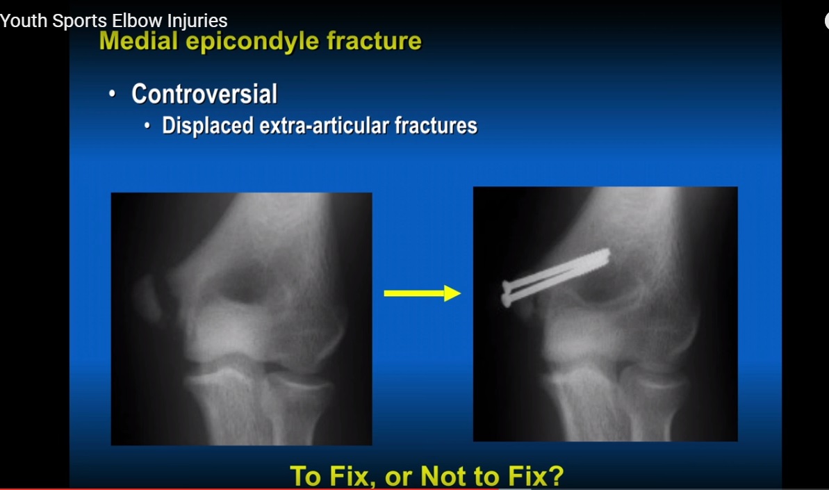
4. Medial Epicondyle Fracture
Anatomy
- Origin of flexor–pronator muscle group
- Ulnar collateral ligament attaches distal to the epicondyle
Mechanism
- Valgus stress
- Avulsion injury
Clinical Importance
- Commonly associated with elbow dislocation
- Fragment may become incarcerated within the joint
Treatment
- Often conservative
Indications for Surgery
- Significant displacement
- Fragment trapped inside joint
- High-demand athlete (throwing arm)
5. Ulnar Collateral Ligament Injury
Mechanism
- Repetitive valgus stress in throwing athletes
Risk Factors
- Excessive pitching duration
- High pitch counts
- High velocity throwing
- Fatigue
Clinical Features
- Medial elbow pain
- Instability
Management
- Rest in early stages
- Reconstruction in severe cases
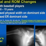
6. Little Leaguer’s Elbow
Definition
- Medial epicondyle apophysitis
Features
- Pain in young throwing athletes
- Widening or fragmentation of apophysis
- Enlargement of dominant side
Treatment
- Rest from throwing
- Gradual return to activity
7. Osteochondritis Dissecans of Capitellum
Mechanism
- Repetitive lateral compartment compression
Features
- Pain with loss of extension
- Mechanical symptoms such as locking
Imaging
- X-ray shows irregular capitellum
- MRI identifies unstable fragments
Treatment
- Early stage: rest
- Advanced stage: arthroscopic debridement with or without microfracture
8. Olecranon Injuries
Types
- Olecranon fracture
- Olecranon apophysitis
Mechanism
- Repetitive traction from triceps
- Common in throwers and gymnasts
Treatment
- Rest
- Surgical fixation if indicated
9. Biomechanics of the Elbow
Forces During Throwing
- Lateral compartment: compression
- Associated with osteochondritis dissecans
- Medial compartment: tension
- Associated with ulnar collateral ligament injury
- Medial epicondyle avulsion
10. Differential Diagnosis by Compartment
Medial
- Medial epicondyle fracture
- Ulnar collateral ligament injury
- Ulnar nerve instability
- Little leaguer’s elbow
Lateral
- Osteochondritis dissecans
- Radial head pathology
- Loose bodies
Posterior
- Olecranon fracture
- Olecranon apophysitis
- Triceps injury
Anterior
- Capsular injury
- Loose bodies
11. Key Exam Pearls
- Most common pediatric elbow fracture: supracondylar fracture
- Most serious complication: Volkmann ischemic contracture
- Always assess pulses and nerve function
- Always obtain post-reduction imaging in elbow dislocation
- Medial elbow pain in throwers suggests UCL injury or apophysitis
- Loss of extension suggests osteochondritis dissecans
- Cubitus varus results from malunion and is mainly cosmetic
12. Prevention
- Limit pitch counts
- Avoid year-round pitching
- Encourage participation in multiple sports
- Early evaluation of elbow pain
Final Takeaway
- Pediatric elbow injuries require:
- Careful neurovascular assessment
- Knowledge of growth plate anatomy
- Awareness of sport-related overuse injuries
- Early recognition prevents:
- Deformity
- Instability
- Long-term dysfunction
Courtesy: Mary Lloyd Ireland M.D. Associate Professor University of Ketucky Lexington, KY, USA www.MaryLloydIreland.com http://orthopaedics.med.uky.edu/
Leave a Reply