
Courtesy: Dr Rishi Kanna, Dr Ashok Shyam, Ortho TV
General Principles
Children are not small adults.
- Anatomy, biomechanics, and injury patterns differ significantly.
- Diagnosis is challenging because of normal anatomical variants, difficult clinical examination, and subtle radiological findings.
Incidence and Distribution
- Pediatric spine injuries account for approximately 1 to 2 percent of pediatric trauma.
- Cervical spine injuries account for about 70 to 75 percent of pediatric spinal injuries.
- Adults have cervical involvement in approximately 30 to 35 percent of cases.
- Children younger than 8 years commonly sustain upper cervical spine injuries (C1 to C3).
- Children older than 8 to 10 years have injury patterns similar to adults.
Anatomical and Radiological Differences
- Synchondroses between ossification centers may mimic fractures.
- Normal anterior vertebral wedging may resemble compression fractures.
- Pseudosubluxation is common, particularly in the cervical spine.
Vertebral Ossification
Each vertebra has three primary ossification centers.
- One centrum.
- Two neural arches.
The 1, 3, 6 rule
- Ossification centers appear at 1 year.
- Neural arches fuse posteriorly by 3 years.
- Neural arches fuse with the vertebral body by 6 to 7 years.
Axis (C2)
- Odontoid synchondrosis fuses at approximately 6 years.
- Can mimic a fracture.
- Represents a weak point prone to injury.
Biomechanics
Children younger than 8 years
- Large head with a small torso.
- Increased ligamentous laxity.
- Horizontal facet joints.
- Fulcrum of motion is in the upper cervical spine.
- Greater risk of instability, neurological injury, and head injury.
Children older than 8 years
- Spine behaves like the adult spine.
- Stronger ligaments.
- Fused synchondroses.
- Fulcrum shifts to the lower cervical spine.
Prehospital and Emergency Care
- Avoid standard flat spine boards in young children.
- Large head causes neck flexion during immobilization.
- Elevate the torso or use an occipital recess to maintain neutral cervical alignment.
Indications for Cervical Spine Imaging
Image the cervical spine in patients with
- Neck pain or tenderness.
- Neurological deficit.
- Altered consciousness.
- Distracting injuries.
- Inconsolable child.
- Unconscious patient.
Pseudosubluxation
- Common at C2 to C3.
- Must be distinguished from true instability.
Swischuk line
- Drawn along the spinolaminar junctions.
- Normal alignment forms a smooth continuous line.
- Disruption suggests true cervical injury.
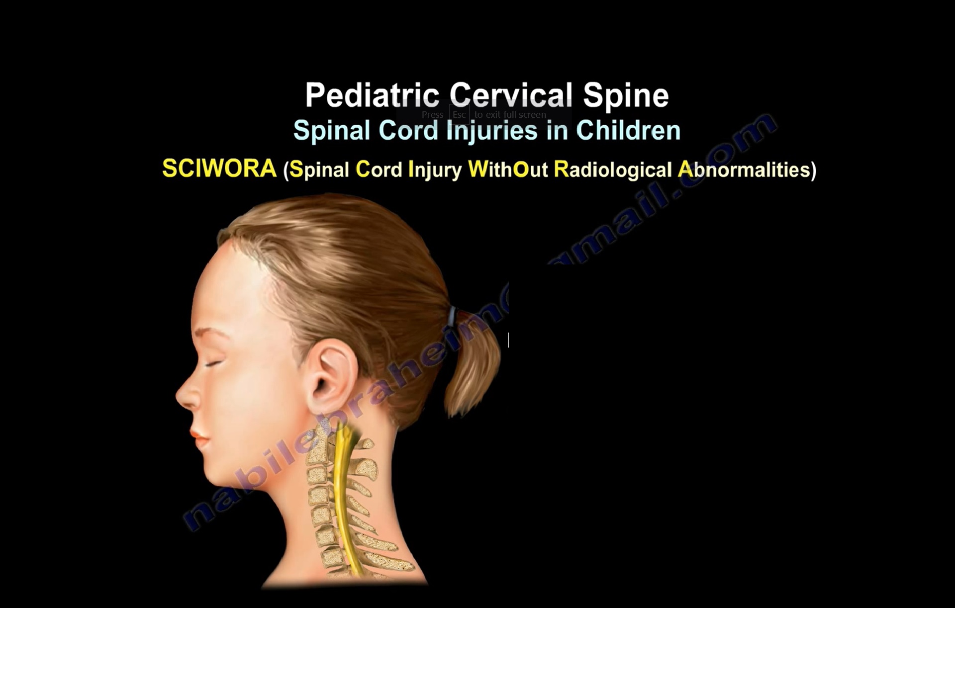
SCIWORA
Spinal Cord Injury Without Radiological Abnormality
- Common in children younger than 8 years.
- Represents 20 to 40 percent of pediatric spinal cord injuries.
Mechanism
- Flexible vertebral column stretches without fracture.
- Less elastic spinal cord sustains injury.
Diagnosis
- MRI is essential.
Management
- Usually nonoperative.
- Prognosis depends on the initial neurological status.
Imaging Strategy
X ray
- First line investigation.
CT scan
- Best for suspected bony injuries.
MRI
- Required when X rays or CT scans are normal but suspicion remains high.
- Essential for diagnosing SCIWORA.
- Evaluates ligaments, soft tissues, and the spinal cord.
Injury Patterns
Children younger than 8 years
- Upper cervical injuries predominate.
- Higher incidence of neurological deficits.
- Higher mortality.
Children older than 10 years
- Adult type fracture and dislocation patterns.
Endplate Avulsion Injury
- Occurs in adolescents after flexion or twisting injuries.
- Can mimic lumbar disc prolapse on MRI.
- CT scan demonstrates a bony endplate avulsion.
Key Clinical Points
- Recognize normal anatomical variants before diagnosing fractures.
- C2 to C3 pseudosubluxation is a common normal finding.
- MRI is essential when neurological deficits are present or diagnosis remains uncertain.
- Management should be individualized based on age, injury pattern, and neurological status.
Exam Pearls
- Pediatric cervical spine injuries are more common than thoracolumbar injuries.
- Upper cervical injuries predominate in children younger than 8 years.
- MRI is the investigation of choice for SCIWORA.
- Swischuk line helps distinguish pseudosubluxation from true cervical instability.
- Odontoid synchondrosis normally fuses at approximately 6 years.
- Large head and ligamentous laxity predispose young children to upper cervical injuries.


Leave a Reply