Courtesy: Prof Nabil Ebraheim, University of Toledo, Ohio, USA
Overview of the Sacrum
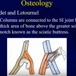
- The sacrum is a large triangular bone located at the base of the vertebral column.
- It forms the posterior part of the pelvis and connects the spine to the pelvic girdle.
- The sacrum articulates superiorly with the fifth lumbar vertebra and laterally with the iliac bones at the sacroiliac joints.
- The bone is broad and thick proximally and becomes thinner and narrower distally.
Posterior Surface of the Sacrum
- The posterior surface contains several important anatomical landmarks.
- The sacroiliac joint lies laterally and may be involved in sacroiliac joint dysfunction and pain.
- Posterior sacral foramina allow passage of the posterior rami of sacral spinal nerves.
- The superior articular processes articulate with the inferior articular processes of the fifth lumbar vertebra.
Sacral Crests
- The median sacral crest represents the fused spinous processes of the sacral vertebrae.
- The intermediate sacral crest represents fused articular processes.
- The lateral sacral crest represents fused transverse processes.
Sacral Canal and Hiatus
- The sacral canal is a continuation of the vertebral canal within the sacrum.
- It transmits sacral nerve roots.
- The sacral hiatus is an opening at the inferior end of the sacral canal.
- It is clinically important for caudal epidural anesthesia.
Sacral Ala and Sacral Tuberosity
- The sacral ala are the wing-like lateral expansions of the upper sacrum.
- They contribute to the formation of the sacroiliac joint.
- The sacral tuberosity is a rough area that provides attachment for ligaments associated with the sacroiliac joint.
Anterior Surface of the Sacrum
- The anterior surface is concave and faces the pelvic cavity.
- It contains anterior sacral foramina which transmit the anterior rami of sacral nerves.
- The sacral promontory forms the anterior projecting margin of the first sacral vertebra.
Superior View of the Sacrum
- The superior view resembles the pelvic inlet seen on radiographic imaging.
- Important structures visible include the sacral ala, body of the sacrum, sacral canal, and superior articular facets.
- The sacroiliac joints are also visible laterally.
Coccyx
- The coccyx lies inferior to the sacrum and forms the terminal segment of the vertebral column.
- It is commonly referred to as the tailbone.
- The coccyx usually consists of two to four fused rudimentary vertebrae.
Functions of the Coccyx
- The coccyx provides attachment sites for ligaments, tendons, and pelvic floor muscles.
- It contributes to pelvic floor stability.
- It acts as a shock absorber when sitting.
Clinical Relevance
- Excessive movement or trauma to the coccyx may cause pain known as coccydynia.
- Sacroiliac joint dysfunction can cause lower back or pelvic pain.
- The sacral hiatus is an important landmark used in caudal epidural injections.

Leave a Reply