
Courtesy: Dr Makridis Konstantinos, Dr Ashok Shyam, Ortho TV
Robotic-Assisted Total Knee Arthroplasty: Kinematic and Restricted Kinematic Alignment (rKA)
Fundamental Biomechanics of Knee Kinematics
-
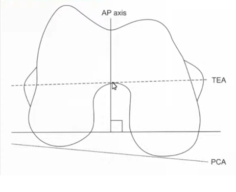
Primary Cylindrical Flexion Axis: The transverse axis of the distal and posterior femoral condyles guides the flexion-extension arc.
-
Asymmetric Native Rollback:
-
During deep flexion, the medial femoral condyle exhibits minimal anteroposterior translation (approximately 2 mm translation).
-
The lateral femoral condyle demonstrates extensive posterior translation and rollback (approximately 20 mm translation).
-
-
Evolution Beyond Equal Rectangular Gaps:
-
Traditional mechanical alignment targeted identical, symmetrical extension and flexion gaps.
-
Contemporary kinematic principles recognize that a physiological knee requires constitutional lateral laxity in flexion to accommodate lateral femoral rollback.
-
-
Restoration of the Three Kinematic Axes: Re-establishing the transverse flexion-extension axis, longitudinal tibial rotation axis, and patellofemoral tracking axis restores native joint balance without excessive soft-tissue releases.
Kinematic Alignment (KA) vs. Restricted Kinematic Alignment (rKA)
-
Kinematic Alignment Premise: Aims to resurface the knee back to its constitutional pre-arthritic anatomy by matching bone resections directly to implant thickness (accounting for cartilage and bone wear).
-
The Clinical Dilemma with Pure KA: Patients with severe constitutional outliers (such as extreme varus or valgus) risk abnormal shear stress on the implant-bone interface and eccentric polyethylene wear.
-
Restricted Kinematic Alignment (rKA) Boundary Philosophy:
-
Designed to safely restore patient-specific anatomy while imposing strict boundaries to protect component longevity.
-
Adjusts extreme bony morphology to keep the overall mechanical alignment and individual bone cuts within a safe envelope.
-
Core Principles & Boundary Parameters of rKA
-
Overall Hip-Knee-Ankle (HKA) Boundary: Maintain the final postoperative mechanical axis within of neutral (177° to 183°).
-
Individual Bone Cut Boundaries:
-
Lateral Distal Femoral Angle (LDFA): Restricted within of perpendicular (typically between 85°–86° and 93°).
-
Medial Proximal Tibial Angle (MPTA): Restricted within of perpendicular (typically between 85°–86° and 93°).
-
-
The “Unworn” vs. “Worn” Resection Principle:
-
On the unworn compartment, resect bone exactly equal to the implant thickness (minus blade kerf).
-
On the worn compartment, compensate for measured cartilage/bone loss to restore the pre-arthritic articular joint line.
-
Prioritize the femoral anatomy first, preserving native femoral condylar geometry to maintain natural kinematics.
-
-
Boundary Adjustments: If initial planned cuts result in an overall limb alignment outside the safe zone, modify the femoral or tibial cuts inward until the construct falls within boundaries.
CPAK Classification and the Arithmetic HKA (aHKA)
-
Arithmetic HKA (aHKA):
-
Calculated as .
-
Represents constitutional limb alignment prior to arthritic joint destruction.
-
Independent of joint space narrowing, subluxation, and distal femoral or proximal tibial cartilage erosion.
-
-
CPAK (Coronal Plane Alignment of the Knee):
-
Matrix incorporating both the constitutional arithmetic axis (varus, neutral, valgus) and joint line obliquity (apex distal, neutral, apex proximal).
-
Guides the surgeon on how far the patient’s native joint deviates from standard neutral mechanical orientation.
-
Role of Robotic Assistance and Navigation
-
Precision in Multiplanar Cuts: Manual instrumentation shows high outlier rates when attempting subtle 1° to 2° angle variations. Robotic platforms execute precise angular corrections reliably.
-
Dynamic Gap Balancing and Soft-Tissue Mapping:
-
Real-time intraoperative tracking of extension and flexion gap laxity throughout the full arc of motion.
-
Quantifies medial stability alongside physiological lateral laxity during flexion without relying solely on subjective manual feel.
-
-
Pre-Planning and Virtual Modification:
-
Allows the surgeon to review bone cuts, component rotation, and predicted limb alignment prior to committing to resections.
-
Enables seamless intraoperative switching between mechanical, kinematic, or restricted kinematic philosophies based on patient anatomy.
-
High-Yield Summary of the rKA Philosophy
-
Preoperative Planning & Robotic Execution: Accurately plan and verify bone cuts to minimize unintended mechanical outliers.
-
Boundary Adherence: Keep individual femoral and tibial cuts within (85°–93°) and overall limb alignment within of neutral.
-
Soft-Tissue Preservation: Restores constitutional joint lines and physiological lateral flexion laxity, substantially decreasing the need for ligament releases.
-
Implant Protection: Protects modern polyethylene inserts and fixation interfaces by filtering out extreme constitutional deformities while preserving the advantages of patient-specific kinematics.

Leave a Reply