
Courtesy: Dr Krishna Kiran, Dr Ashok Shyam, Ortho TV
Knee Kinematics and Total Knee Arthroplasty Alignment Philosophies
Native Knee Kinematic Anatomy
-
Asymmetric Joint Morphology: Medial and lateral femoral condyles and tibial plateaus are asymmetric.
-
Meniscal Constraints:
-
The medial meniscus is firmly anchored to the deep medial collateral ligament (MCL).
-
The lateral meniscus is mobile, sliding posteriorly during flexion.
-
-
Four-Bar Linkage: The anterior cruciate ligament (ACL) and posterior cruciate ligament (PCL) guide physiological motion.
-
Normal Knee Motion:
-
Displays lateral pivoting in extension.
-
Displays medial pivoting in flexion.
-
-
Deep Flexion Mechanism: The lateral femoral condyle subluxates posteriorly beyond 140° of flexion as the lateral meniscus translates.
-
Prosthetic Kinematic Discrepancy:
-
Cruciate-retaining (CR) knees often show paradoxical anterior femoral slide.
-
Posterior-stabilized (PS) designs achieve only 60% to 70% of native femoral rollback via cam-post engagement.
-
Current total knee arthroplasty (TKA) designs cannot fully duplicate native knee kinematics.
-
Unicompartmental knee arthroplasty (UKA) remains closest to native kinematics because both cruciates and the lateral meniscus remain intact.
-
Constitutional Varus and Alignment Debates
-
Historical Threshold: Traditional mechanical alignment targeted a neutral Hip-Knee-Ankle (HKA) axis within .
-
Constitutional Varus:
-
Bellemans et al. found that 32% of asymptomatic men and 17% of asymptomatic women naturally have constitutional varus greater than 3°.
-
Original criteria relied on short-leg radiographs rather than full-length weight-bearing films.
-
-
Uncertain Patient Satisfaction Factors:
-
Unhappy TKA patients may stem from incorrect initial indications, low pain thresholds, or psychosocial variables.
-
Altering alignment alone does not reliably resolve patient dissatisfaction.
-
Personalized Alignment Strategies
-
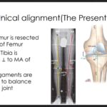
Kinematic Alignment (KA): Resects bone to match native pre-arthritic anatomy without soft-tissue releases.
-
Inverse Kinematic Alignment (iKA): Reconstructs the joint starting from the native tibial joint line.
-
Restricted Kinematic Alignment (rKA): Re-creates patient anatomy within safe boundaries (usually ).
-
Functional Alignment (FA): Adjusts implant orientation within planned boundaries to balance intraoperative ligament gaps.
Population Anatomy & The Vendittoli Data
-
Vendittoli Findings:
-
Only 50% of the population presents with a neutral HKA axis where a lateral distal femoral angle (LDFA) of 3° balances a medial proximal tibial angle (MPTA) of 3°.
-
50% of the population are constitutional outliers.
-
Widening safe alignment boundaries to captures 80% of the population.
-
-
Indian Population Data: Normative population distributions for MPTA and LDFA in Indian cohorts remain undefined.
-
CPAK Classification: Coronal Plane Alignment of the Knee groups knees based on constitutional arithmetic HKA and joint line obliquity.
Biomechanics: Polyethylene Constraints and Pressures
-
Polyethylene Limitations: Replicating extreme native varus or valgus anatomy risks eccentric edge loading, early polyethylene wear, and component loosening.
-
Intercompartmental Pressures:
-
Kinematic alignment balances medial and lateral compartment pressures more evenly than mechanical alignment.
-
Conventional mechanical techniques rely heavily on subjective tactile feel rather than quantitative load measurements.
-
-
Plane Alignment Accuracy:
-
Manual instrumentation poorly controls sagittal and rotational alignments.
-
3D preoperative planning and intraoperative navigation or robotics are required to accurately control multiplanar cuts.
-
Clinical Outcomes: Mechanical vs. Kinematic Alignment
-
Long-Term Survivorship:
-
Stephen Howell et al. demonstrated comparable 10-year clinical outcomes for kinematic alignment.
-
Registry databases show identical revision rates of roughly 3% at 10 years for both mechanical and kinematic alignment.
-
-
Clinical Equivalence: Dr. Ashok Rajgopal noted that altering component alignment by 2° to 3° does not reliably produce clinically meaningful functional gains.
-
Modern Surgical Realignment: The main benefit of personalized alignment has been minimizing extensive soft-tissue releases during balancing.
-
Alignment Philosophies Summary:
-
Kinematic alignment starts from native joint morphology.
-
Mechanical alignment starts from soft-tissue releases to fit neutral bone cuts.
-
Functional and restricted kinematic alignments balance native anatomy within set implant limits.
-
-
Current Standard: Mechanical alignment remains the global gold standard until dual-cruciate-retaining, motion-guided implants are refined.



Leave a Reply