
Courtesy: Kaye Wilkins MD
Prof Lynn Staheli
Radial Neck and Olecranon Fractures in Children
Radial Neck Fractures
Basic concepts
- Most pediatric radial fractures occur at the radial neck (metaphysis), the weakest part of the proximal radius.
- Radial head ossification appears at 3 to 4 years of age.
- Ossification is complete by 6 to 7 years.
- A bipartite radial head may mimic a fracture.
Biomechanics
- The radiocapitellar joint is highly congruent.
- Translocation of the radial head produces a cam effect, limiting forearm rotation, particularly pronation.
- Even minimal displacement can significantly impair function.
Mechanism of injury
Primary injuries
- Fall on an outstretched hand.
- Elbow extension with valgus stress causing compression of the radial head against the capitellum.
Associated injuries
- Monteggia fracture dislocation, particularly Type II.
- Elbow dislocation.
Patterns of displacement
- Valgus compression causing angulation with or without translocation.
- Elbow dislocation may produce anterior displacement initially and posterior displacement after reduction.
Assessment
- Measure fracture angulation.
- Assess radial head translocation.
- Examine forearm pronation and supination.
- Functional motion requires approximately 50 degrees each of pronation and supination.
Acceptable limits
- Angulation less than 30 degrees is acceptable.
- Angulation between 30 and 60 degrees usually requires closed reduction.
- Angulation greater than 60 degrees often requires surgical treatment.
- Translocation less than 3 mm is generally acceptable.
- Greater translocation should be corrected to preserve rotation.
Treatment
Nonoperative treatment
- Immobilization for minimally displaced fractures.
Closed reduction
- Israeli (Kaufman) technique.
- Elbow flexed to 90 degrees.
- Direct pressure over the radial head.
- Forearm pronation helps close the metaphyseal gap and reduce the fragment.
Percutaneous techniques
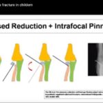
- Direct reduction using a K wire or elevator.
- Retrograde intramedullary nailing (Metaizeau technique).
- Rotation of the intramedullary nail assists fracture reduction.
Open reduction
- Reserved for failed closed or percutaneous reduction.
- Indicated for displaced Salter Harris Type III fractures.
- Higher risk of complications.
Complications
- Loss of forearm rotation.
- Avascular necrosis.
- Malunion.
- Nonunion.
- Radial head overgrowth.
- Radioulnar synostosis.
- Heterotopic ossification.
Clinical pearls
- Always evaluate the ulna for a Monteggia injury.
- Assess the lateral collateral ligament complex.
- Severe displacement increases the risk of avascular necrosis.
- Look carefully for associated injuries.
Olecranon Fractures
Ossification
- Olecranon ossification begins at approximately 8 years of age.
- Two ossification centers are present: articular and traction centers.
Mechanism based classification
Flexion (tension) injury
- Triceps pull causes posterior displacement.
- Associated with loss of active elbow extension.
- Posterior swelling and a palpable gap may be present.
Extension injury
- Failure of the anterior cortex with an intact posterior cortex.
- Stable injury suitable for immobilization in elbow flexion.
Shear injury
- Caused by direct trauma to a flexed elbow.
- Radius and ulna displace together.
Greenstick injury
- May be associated with radial neck fractures.
- May occur with medial epicondyle fractures.
- May accompany Monteggia lesions.
Treatment
Nonoperative treatment
- Long arm cast for minimally displaced fractures.
Operative treatment
- Restore the articular surface.
- Restore the extensor mechanism.
- Tension band wiring.
- Screw fixation with tension band augmentation.
- Absorbable suture fixation may be advantageous in children.
Complications
- Loss of elbow extension.
- Prominent hardware.
- Nonunion.
- Residual deformity affecting radiocapitellar alignment.
Exam Pearls
- Radial neck fractures are typically valgus compression injuries.
- Evaluate both angulation and translocation when planning treatment.
- The Israeli (Kaufman) technique is a commonly tested closed reduction method.
- In Monteggia injuries, reduce and stabilize the ulna first.
- Open reduction of radial neck fractures has the highest complication rate.
- Flexion type olecranon fractures commonly present with loss of active elbow extension.
- Tension band fixation converts tensile forces into compressive forces across the fracture site.

Leave a Reply