Courtesy: Prof Lyndon Mason, FRCSOrth, Liverpool, UK
Background
Historically, posterior malleolar fractures were treated according to the “one third rule”:
- Fix the fragment if it involves more than 25 to 30% of the articular surface.
- Leave smaller fragments untreated.
Modern evidence has shown that:
- Fragment size alone does not determine outcome.
- Fracture morphology is more important than size.
- Anatomical reduction of the articular surface is the major determinant of outcome.
Goals of Treatment
The aims of treatment are:
- Anatomical reduction of the articular surface
- Restoration of ankle stability
- Stable fixation
- Prevention of post traumatic arthritis
- Early mobilization
Key principle:
Reduction quality is more important than fragment size.
Importance of CT Scan
CT scan is mandatory for proper evaluation.
Why CT is Important
- Plain radiographs have only about 22% diagnostic accuracy.
- CT frequently changes surgical planning.
- Defines fracture morphology accurately.
- Identifies posteromedial fragments.
- Detects impacted articular fragments.
- Demonstrates soft tissue entrapment.
Clinical Pearl
Every posterior malleolar fracture should undergo CT evaluation before definitive treatment.
Soft Tissue Entrapment
Posterior malleolar fractures may trap soft tissues, particularly:
- Tibialis posterior tendon
Risk Factors
- Fracture line extending into the tibialis posterior tendon sheath.
Reported Risks
- Minor tendon entrapment: approximately 34%
- Major tendon entrapment: approximately 7%
Failure to identify tendon incarceration may result in:
- Malreduction
- Persistent pain
- Poor functional outcome
Mechanism of Injury
1. Ligamentous Avulsion Injury
Mechanism:
- Avulsion by the Posterior Inferior Tibiofibular Ligament (PITFL)
- Foot relatively unloaded
Produces:
- Small posterior fragment
2. Rotational Pilon Injury
Mechanism:
- Supination external rotation injury
- Talus impacts the posterior tibial plafond during rotation
Produces:
- Posterolateral or posteromedial fragments
3. Axial Posterior Pilon Injury
Mechanism:
- Axial load through a plantarflexed ankle
Produces:
- Large posterior articular fragment
- Significant joint impaction
Mason Molloy Classification
Type 1
PITFL Avulsion Fracture
Characteristics:
- Small fragment
- Ligament avulsion injury
- No significant talar impaction
Type 2A
Rotational Pilon Fracture
Characteristics:
- Isolated posterolateral fragment
Type 2B
Rotational Pilon Fracture
Characteristics:
- Posterolateral fragment
- Additional posteromedial fragment
Important point:
- Posteromedial fragment requires separate reduction and fixation.
Type 3
Axial Posterior Pilon Fracture
Characteristics:
- Large posterior fragment
- Significant articular injury
- Caused by axial loading
Posterior Malleolus and Rotational Stability
Traditional teaching focused on:
- Prevention of posterior talar translation
Modern studies show that the posterior malleolus is critical for:
- Rotational stability of the ankle
- Syndesmotic stability
Even fragments involving less than 25% of the articular surface can significantly affect rotational stability.
Fragment Size Versus Outcome
Current evidence demonstrates:
Fragment Size
- Poor correlation with clinical outcome
Articular Reduction
- Strong correlation with clinical outcome
Poor results occur with:
- Residual step off
- Articular incongruity
- Malreduction
- Inadequate fixation
Syndesmotic Stability
The posterior malleolus forms an important attachment of the PITFL.
Fixation of the posterior fragment may:
- Restore syndesmotic stability
- Reduce need for syndesmotic screws
However:
- Not every posterior malleolar fracture stabilizes the syndesmosis completely.
- High fibular fractures and severe syndesmotic injuries may still require syndesmotic fixation.
Surgical Approaches
Posterolateral Approach
Structures at Risk
- Sural nerve
- Peroneal vessels
Advantages
- Familiar approach
- Good access to posterolateral fragments
Limitations
- Limited visualization
- Difficult access to posteromedial fragments
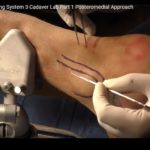
Posteromedial Approach
Currently considered the workhorse approach.
Surgical Interval
Between:
- Tibialis posterior
- Flexor digitorum longus
Advantages
- Excellent exposure
- Direct visualization
- Access to both posterolateral and posteromedial fragments
- Easier reduction of complex fracture patterns
Posteromedial Extension
Provides access to:
- Large posterior pilon fragments
- Die punch fragments
- Impacted articular segments
Patient Positioning
Prone Position
Traditional method.
Semi Prone (Recovery Position)
Advantages:
- Easier anesthesia management
- Better fluoroscopic imaging
- Improved access to medial and posterior ankle
- Easier repositioning if necessary
Order of Fixation
For Type 2B and complex posterior fractures:
Step 1
Fix the posteromedial fragment first.
Step 2
Fix the posterolateral fragment.
Step 3
Fix the fibula.
Step 4
Assess syndesmotic stability.
Important Pearl
If the posterolateral fragment is fixed first, the posteromedial fragment may displace or “spit out.”
Die Punch Fragments
Definition:
- Impacted osteochondral fragments within the articular surface.
Importance:
- Associated with worse outcomes.
- Frequently missed on fluoroscopy.
Best managed by:
- Direct visualization
- Careful CT planning
- Anatomical reduction
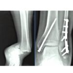
Fragment Specific Fixation
Modern philosophy emphasizes:
- Individual reduction of each fragment
- Separate fixation when necessary
Benefits:
- Improved anatomical reduction
- Better restoration of joint congruity
- Lower incidence of post traumatic arthritis
- Better functional outcomes
Direct Versus Indirect Fixation
Indirect Fixation
Traditional method:
- Anterior to posterior screw fixation
Limitations:
- No direct visualization
- Higher risk of malreduction
Direct Fixation
Preferred modern technique.
Advantages:
- Direct visualization
- Accurate reduction
- Better fixation
- Improved outcomes
Indirect fixation may still be acceptable if:
- Arthroscopy confirms reduction, or
- Intraoperative CT confirms alignment.
Safe Zone for Screw Placement
Avoid:
- Penetration into the fibular incisura
Incorrect screw placement may cause:
- Syndesmotic malreduction
- Joint incongruity
- Persistent instability
Key Examination Pearls
- CT scan is essential in all posterior malleolar fractures.
- Fracture morphology is more important than fragment size.
- Posterior malleolus is a major stabilizer of ankle rotation and syndesmosis.
- Articular reduction determines outcome.
- Posteromedial fragments require independent assessment and fixation.
- Posteromedial approach provides superior visualization in complex fractures.
- Direct reduction and fragment specific fixation represent the current standard of care.
Leave a Reply