
Courtesy: Prof Nabil Ebraheim, University of Toledo, Ohio, USA
Physical Examination of the Lower Spine and Lower Extremity
Overview
The examination follows the usual orthopaedic sequence:
Inspection – Palpation – Range of motion – Muscle strength – Provocative/special tests -Thorough neurovascular examination
The neurological component should assess motor, sensory, reflexes, and upper motor neuron signs where relevant.
- Inspection
Deformity and Alignment
Inspect for deformity in both:
- Coronal plane
- Scoliosis
- Pelvic obliquity
- Sagittal plane
- Cervical lordosis
- Thoracic kyphosis
- Lumbar lordosis
Also inspect the patient from both front and back for asymmetry.
Skin and Soft Tissue
Look for:
- Café-au-lait spots
- Hairy patches
- Dimples
- Previous surgical scars
- Muscle wasting
specifically states that calf muscle atrophy may suggest L5–S1 weakness or neuropathy.
- Palpation
Palpate the important bony landmarks:
- Iliac crests
- Posterior superior iliac spines
- Spinous processes
- Sacrum
- Greater trochanters
- Ischial tuberosities
Also palpate soft tissues for tenderness or trigger points involving:
- Paravertebral muscles
- Gluteal muscles
- Piriformis
- Sciatic notch
- Sacroiliac joints
also mentions attempting to palpate the sciatic nerve.
- Gait Examination
Gait gives important clues to pathology.
| Gait abnormality | Possible implication in transcript |
| Antalgic gait | Hip arthritis |
| Trendelenburg gait | Hip abductor weakness |
| Steppage gait | Foot drop / ankle dorsiflexor weakness |
| Wide-based staggering gait | Cervical myelopathy / neurological disorder |
An important point is that cervical myelopathy can coexist with lumbar pathology, so gait disturbance should not automatically be attributed to the lumbar spine.
- Pain With Spinal Movement
stresses asking:
Which movement causes pain, and which movement relieves it
Lumbar Spinal Stenosis
- Extension increases pain
- Flexion improves symptoms
Disc Pathology
- Flexion increases pain
- Sitting and leaning forward may increase disc-related pain according to
Therefore, presents these as opposite patterns:
Stenosis – worse with extension, better with flexion
Disc pathology – worse with flexion
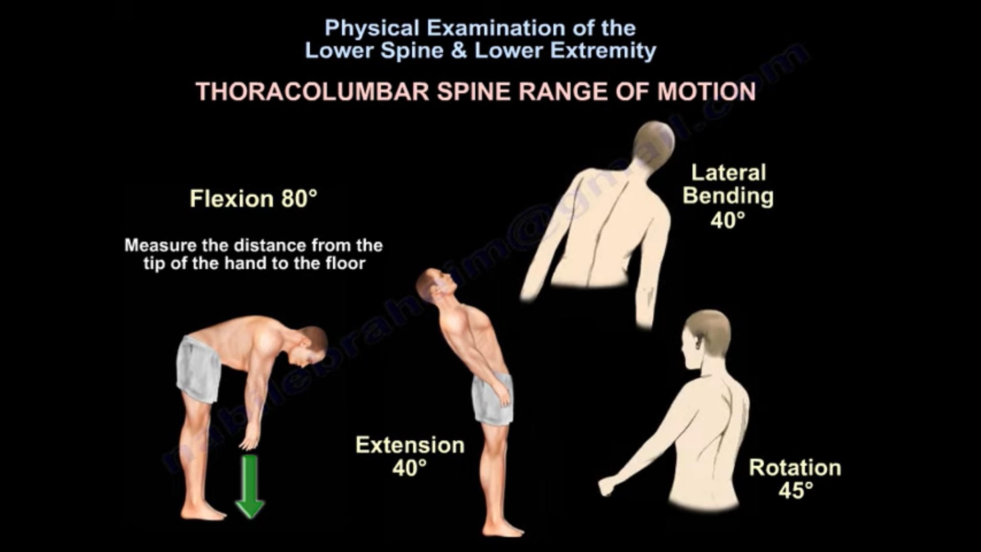
- Thoracolumbar Range of Motion
gives approximate values:
| Movement | ROM stated |
| Flexion | ~80° |
| Extension | ~40° |
| Lateral bending | ~40° |
| Rotation | ~45° |
For flexion, the distance from the fingertips to the floor may also be assessed.
notes that these values can vary.
- Neurological Examination
Assess individual nerve roots from approximately:
L2 to S1
The examination includes:
- Sensory testing
- Motor testing
- Reflexes
- Provocative tests
- Upper motor neuron signs when indicated
- Sensory Examination
describes testing four sensory modalities:
- Pain
- Light touch
- Temperature
- Proprioception
In routine practice, says pain and light touch are most commonly tested.
The aim is to identify:
- A dermatomal pattern – suggests nerve-root pathology
- A glove/stocking type distribution – may suggest peripheral neuropathy
Dermatomal Distribution
| Root | Sensory area |
| L2 | Mid-anterior thigh |
| L3 | Distal thigh / knee area |
| L4 | Medial leg to medial foot |
| L5 | Dorsum of leg/foot |
| S1 | Lateral and plantar aspect of foot |
- Motor Examination
Hip Flexion
Main muscle:
Iliopsoas
Roots stated:
L1, L2, L3
Hip Adduction
Roots:
L2, L3, L4
Knee Extension
Roots:
L2, L3, L4
emphasizes L4 as an important contributor.
Ankle Dorsiflexion
Main muscle:
Tibialis anterior
Roots stated:
L4–L5
L5
Commonly tested by:
- Great toe extension
- Hip abduction via gluteus medius
S1
Commonly tested by:
Ankle plantar flexion
Main muscle group:
Gastrocnemius–soleus complex
High-Yield Motor Root Table
| Root | Key motor test |
| L2 | Hip flexion |
| L3 | Knee extension |
| L4 | Ankle dorsiflexion / knee extension |
| L5 | Great toe extension / hip abduction |
| S1 | Plantar flexion |
- Reflexes
mentions two main lower-extremity reflexes:
- Patellar reflex
- Achilles tendon reflex
These are used as part of the lower-limb neurological examination.
- Provocative Tests
lists:
- Straight leg raise
- Contralateral straight leg raise
- Femoral stretch test
- Clonus
- Babinski
Straight Leg Raise
Used for:
L5–S1 nerve-root irritation
Contralateral SLR
associates this with:
Sequestered disc causing L5/S1 root irritation
Femoral Stretch Test
Used for:
L3–L4 nerve-root irritation
- Upper Motor Neuron Signs
mentions:
- Clonus
- Babinski
These are used to look for an upper motor neuron lesion.
This is particularly relevant when cervical cord pathology may coexist with lumbar complaints.
- Bulbocavernosus Reflex
discusses the bulbocavernosus reflex in relation to spinal shock.
The maneuver is described using rectal/urethral/genital stimulation, with a positive response consisting of:
Anal sphincter contraction
According to , return of this reflex indicates:
End of spinal shock
- Sacroiliac Joint Examination
FABER Test
FABER is discussed as a useful SI-joint test, but states:
It is not confirmatory.
Another useful historical/localizing feature is:
The patient pointing with one finger directly to the posterior SI joint as the site of pain.
considers this almost as useful as the FABER maneuver.
Diagnostic SI Injection
When SI-joint pain is suspected, suggests confirmation by:
Injection of local anesthetic into the SI joint followed by pain relief.
- Waddell Signs
Waddell signs are described as:
Non-organic physical examination findings
Examples mentioned include:
- Non-anatomic/inconsistent weakness
- Superficial or non-anatomic tenderness
- Findings disproportionate to the rest of the examination
states that three or more positive signs may suggest that the complaints do not fit a purely anatomical pattern, but also emphasizes that:
Waddell signs are controversial.
They should therefore not be used simplistically to label a patient.
High-Yield Differential Pattern
| Finding | Favors |
| Pain worse with lumbar extension | Lumbar stenosis |
| Pain better with flexion | Lumbar stenosis |
| Pain worse with flexion | Disc pathology |
| Antalgic gait | Hip pathology |
| Trendelenburg gait | Hip abductor weakness |
| Steppage gait | Foot drop |
| Wide-based gait | Myelopathy |
| SLR positive | L5/S1 root irritation |
| Femoral stretch positive | L3/L4 root irritation |
| Clonus/Babinski | UMN lesion |
| Finger pointing to SI joint | SI-joint source |
Practical Examination Sequence
INSPECTION
Coronal deformity – sagittal alignment – pelvic obliquity -skin – scars -wasting
PALPATION
Iliac crest – PSIS – spinous processes – sacrum – greater trochanter – ischial tuberosity – soft tissues
GAIT
Antalgic – Trendelenburg – steppage- wide based
ROM
Flexion – extension – lateral bending – rotation
MOTOR
L2 – L3 – L4 – L5 – S1
SENSORY
L2 – L3 – L4 – L5 -S1
REFLEXES
PROVOCATIVE TESTS
SLR – crossed SLR – femoral stretch
UMN SIGNS
Clonus – Babinski
SI JOINT / NON-ORGANIC SIGNS
FABER – SI localization – Waddell signs
Key Take-Home Points
- Lower-spine examination should remain systematic and bilateral.
- Always inspect the entire spine in both coronal and sagittal planes.
- Gait can reveal hip, neurological, dorsiflexor, and cord pathology.
- Extension-provoked pain favors lumbar stenosis, whereas flexion-provoked pain favors disc pathology in ‘s framework.
- Sensory testing should look for a dermatomal distribution.
- Motor examination from L2 to S1 is essential.
- L5 – great toe extension and S1 – plantar flexion are particularly useful quick tests.
- SLR evaluates lower lumbar/sacral root irritation; femoral stretch evaluates higher lumbar roots.
- Do not forget clonus and Babinski when cord disease is possible.
- FABER may support SI-joint pathology, but does not consider it confirmatory.
- Waddell signs are controversial and should be interpreted cautiously.
Exam Pearls
L2 – Hip flexion
L3 – Knee extension
L4 – Ankle dorsiflexion
L5 – Great toe extension
S1 – Plantar flexion
SLR – L5/S1
Femoral stretch – L3/L4
Stenosis – extension hurts, flexion relieves
Disc – flexion hurts
Clonus/Babinski – UMN lesion
Leave a Reply