Courtesy Dr Jefferey Yao, Dr Ashok Shyam, Ortho TV
Overview
Wrist ligaments function as static stabilizers, guiding and constraining motion of the carpal bones.
Types of Wrist Ligaments
1. Intrinsic Ligaments
- Located entirely within the carpus
- Connect adjacent carpal bones
Examples:
- Scapholunate ligament
- Lunotriquetral ligament
2. Extrinsic Ligaments
- Connect carpal bones to the radius or ulna
- Cross:
- Radiocarpal joint
- Midcarpal joint
Concept of Carpal Instability
Important Principle
Malalignment — Instability
- Some individuals may show radiographic malalignment but remain asymptomatic
True Carpal Instability
Defined as:
- Abnormal load transfer
- Abnormal intercarpal motion
- Associated pain and functional impairment
Classification of Carpal Instability (Mayfield)
1. Carpal Instability Dissociative (CID)
- Instability within a carpal row
- Example: Scapholunate dissociation
2. Carpal Instability Non-Dissociative (CIND)
- Instability between proximal and distal rows
3. Carpal Instability Complex (CIC)
- Combination of CID and CIND
- Seen in perilunate dislocations
Perilunate Injuries
Overview
- Represent a spectrum of high-energy wrist injuries
- Frequently missed on initial radiographs
Common Mechanisms
- Motorcycle accidents
- Falls from height
- Falls from ladders/scaffolding
Mechanism of Injury
- Wrist hyperextension
- Ulnar deviation
- Carpal supination
Mayfield Classification of Perilunate Instability
Stage I
- Scapholunate ligament rupture
- May include scaphoid fracture (greater arc)
Stage II
- Injury extends to capitolunate joint
- May involve:
- Capitate dislocation
- Capitate fracture
Stage III
- Lunotriquetral ligament injury
- Lunate remains in fossa
Stage IV
- Complete lunate dislocation
- Lunate displaced volarly into carpal tunnel
Types of Perilunate Injuries
Lesser Arc Injuries
- Pure ligamentous injuries
- No fractures
Greater Arc Injuries
- Associated fractures:
- Scaphoid
- Capitate
- Triquetrum
- Radial styloid
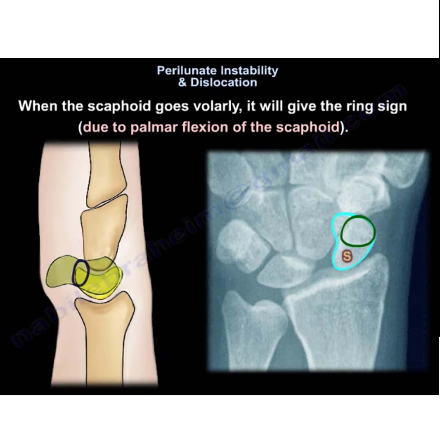
Radiographic Diagnosis
PA View
- Loss of Gilula’s arcs
- Triangular lunate — “Piece-of-pie sign”
Lateral View
- Volar lunate displacement
- “Spilled teacup sign”
Reverse Perilunate Injury
- Begins on ulnar side
- Starts with lunotriquetral ligament injury
- Progresses toward scapholunate region
Initial Management
Median Nerve Assessment
- Compression common due to lunate displacement
Emergency Closed Reduction
Steps
- Apply longitudinal traction
- Hyperextend wrist
- Stabilize lunate with thumb
- Gradually flex wrist
- Reduce capitate into alignment
Definitive Treatment
General Principle
Most cases require early surgical stabilization
1. Arthroscopic Management
Procedures
- Reduction
- Percutaneous pinning
- Capsular repair
Advantages
- Less soft tissue damage
- Preserves blood supply
- Reduced scarring
- Allows assessment of associated injuries
2. Open Reduction and Internal Fixation (ORIF)
Dorsal Approach
- Access to scapholunate ligament
- Key stabilizing structure
Volar Approach
- Access to:
- Lunotriquetral ligament
- Volar capsule
- Allows median nerve decompression
Combined Approach
- Used in complex injuries
3. Arthroscopic Ligament Repair
- Suture-based repair
- Minimally invasive
- Effective stabilization
Management of Advanced Injuries
Stage III & IV Injuries
- Open reduction
- Ligament repair (suture anchors)
- Temporary fixation (K-wires or screws)
Temporary Screw Fixation
Advantages
- Allows early mobilization
- Screws removed after ~3 months
Perilunate Fracture Dislocations
Examples and Treatment
Trans-Scaphoid
- Scaphoid fixation + ligament repair
Trans-Capitate
- Capitate fixation + stabilization
Trans-Radial Styloid
- Radial styloid fixation + carpal alignment
Complications
Early Complications
Missed Diagnosis
- Most common complication
Median Neuropathy
- Often improves after reduction
- Persistent symptoms ? urgent decompression
Avascular Necrosis (AVN)
- May affect:
- Scaphoid
- Lunate
- Less common if ligaments preserved
Late Complications
Chronic Carpal Instability
- Scapholunate instability
Post-Traumatic Arthritis
- Occurs in >50% long-term
Salvage Procedures
Indications
- Chronic or missed injuries
Options
- Proximal row carpectomy
- Limited wrist fusion
- Total wrist arthrodesis
Key Take-Home Points
- Perilunate injuries are high-energy and frequently missed
- Careful evaluation of:
- Gilula’s lines
- Lunate alignment
Management Principles
- Urgent closed reduction
- Assess median nerve
- Early surgical stabilization
Clinical Insight
- Delay >3 months ? often requires salvage procedures

Leave a Reply