Courtesy: Prof Nabil Ebraheim, University of Toledo, Ohio, USA
Paget Disease of the Spine & Differential Diagnosis of Vertebral Sclerosis
Overview
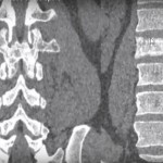
When evaluating lumbar spine X-rays, a vertebra may appear abnormally sclerotic (white).
This finding requires careful differentiation between multiple conditions, as imaging alone may not be sufficient.
Common Differential Diagnoses
- Paget disease of bone
- Prostate cancer metastasis
- Breast cancer metastasis
- Hyperparathyroidism
Important Clinical Note
- Diagnosis often requires:
- Clinical correlation
- Laboratory investigations
- Occasionally biopsy
Paget Disease of Bone (Spinal Involvement)
Radiological Features
Classic Appearance: “Picture Frame Vertebra”
- Thickened cortical margins
- Vertebral body expansion
- Coarsened trabecular pattern
- Increased cortical density at edges
Cortex outlines the vertebra like a picture frame
Pathophysiology
- Initial phase:
- Increased osteoclastic bone resorption
- Followed by:
- Disorganized osteoblastic bone formation
Results in structurally weak, abnormal bone
Laboratory Findings
- Elevated serum alkaline phosphatase
- Increased bone turnover markers:
- Urinary hydroxyproline
- Collagen breakdown products
Histological Features
- Mosaic pattern of lamellar bone
- Prominent cement lines
- Cortical thickening
Additional Features
- May be polyostotic
- Vertebra shows:
- Trabecular coarsening (not uniform sclerosis)
Metastatic Prostate Cancer to the Spine
Overview
- Common cause of osteoblastic (sclerotic) metastases
- Typically affects:
- Lumbar spine
- Elderly men
Radiological Feature: “Ivory Vertebra”
- Diffuse, homogeneous sclerosis
- Vertebral size remains normal
- Disc spaces preserved
Diagnostic Clues
- Elevated PSA (Prostate-Specific Antigen)
- Histology:
- Adenocarcinoma with gland formation
Other Tumors
- Breast cancer may also produce:
- Sclerotic metastases
Rugger-Jersey Spine (Hyperparathyroidism)
Definition
- Classic radiological sign seen in:
- Secondary hyperparathyroidism
Radiological Features
- Dense sclerosis of:
- Superior endplate
- Inferior endplate
- Central vertebral body:
- Relatively lucent
Appearance
- Alternating dense and lucent bands
Resembles rugby jersey stripes
Pathophysiology
- Excess parathyroid hormone (PTH) causes:
- Increased bone resorption
- Loss of bone mass
- Osteoid formation with poor mineralization
Key Radiological Differences
| Condition | Radiological Appearance | Vertebral Size | Key Clues |
|---|---|---|---|
| Paget disease | Picture-frame vertebra, coarse trabeculae | Expanded | High ALP |
| Prostate metastasis | Ivory vertebra (uniform sclerosis) | Normal | High PSA |
| Hyperparathyroidism | Rugger-jersey spine | Normal | High PTH |
Clinical Pearls
- Uniformly sclerotic vertebra in elderly male
–Suspect prostate metastasis until proven otherwise
- Expanded vertebra with cortical thickening + coarse trabeculae
–Suggests Paget disease
- Band-like sclerosis at endplates
— Suggests hyperparathyroidism
Key Takeaways
- Vertebral sclerosis has multiple important differentials
- Imaging findings must be interpreted with:
- Clinical history
- Laboratory markers
- Paget disease shows:
- Expansion + cortical thickening
- Prostate metastasis shows:
- Uniform dense vertebra without expansion


Leave a Reply