Courtesy: Ajith Appuhamy, FRCS Tr and Orth, Srilanka
Paget Disease of Bone
Overview
Paget disease of bone is a chronic metabolic bone disorder characterized by abnormal bone remodeling.
Key Pathological Process
- Excessive osteoclastic bone resorption
- Followed by disorganized osteoblastic bone formation
Resulting Bone Characteristics
- Structurally weak
- Hypervascular
- Deformed
Leads to altered joint biomechanics and secondary osteoarthritis
Types of Paget Disease
Polyostotic Disease (80–85%)
- Involves multiple bones
Monostotic Disease (15–20%)
- Involves a single bone
Epidemiology
- More common after 40 years of age
- Prevalence increases with age
Geographical Distribution
- Higher prevalence:
- United Kingdom
- Australia
- New Zealand
- Less common:
- Asian populations
Etiology
1. Viral Hypothesis
- Possible involvement of:
- Measles virus
- Respiratory syncytial virus
- Canine distemper virus
2. Genetic Factors
- Familial cases: 5–40%
- First-degree relatives may be affected
3. Environmental Factors
- Possible but not clearly established
Pathophysiology
Three Phases of Disease
1. Osteolytic Phase
- Increased osteoclast activity
- Excess bone resorption
2. Mixed Phase
- Simultaneous:
- Resorption
- Formation
3. Sclerotic (Burnt-Out) Phase
- Predominant osteoblastic activity
- Dense, sclerotic bone formation
Key Feature
- Disorganized mosaic pattern of bone
Clinical Manifestations
Orthopedic Features
- Bone pain
- Secondary osteoarthritis
- Pathological fractures
- Bone deformities
Common Deformities
- Bowing of long bones
- Spinal deformities
Non-Orthopedic Features
- High-output cardiac failure (due to hypervascular bone)
- Cranial nerve compression
- Hearing loss
- Raised intracranial pressure
- Neurological deficits
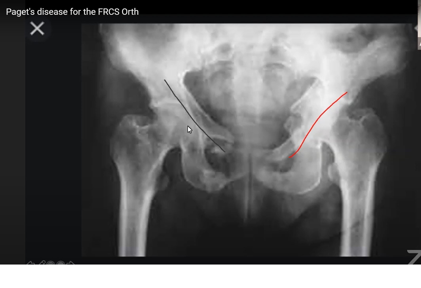
Radiological Features
Pelvis
- Thickened trabeculae
- Cortical thickening
- Pelvic brim sign
- Protrusio acetabuli
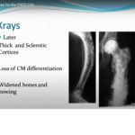
Long Bones
- Cortical thickening
- Candle flame–shaped lytic lesions
- Bowing deformity
- Coarse trabeculae
Skull
- Enlargement
- Early:
- Osteoporosis circumscripta
- Late:
- Cotton wool appearance
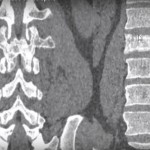
Spine
- Enlarged vertebra
- Picture-frame vertebra
- Late stage:
- Ivory vertebra
Laboratory Findings
Typical Findings
- Elevated alkaline phosphatase
- Increased bone turnover markers:
- Urinary hydroxyproline
- N-telopeptide
- C-telopeptide
- Deoxypyridinoline
Important Note
- Serum calcium — Normal
- Serum phosphate — Normal
Complications
Skeletal
- Bone pain
- Osteoarthritis
- Pathological fractures
- Spinal stenosis
Neurological
- Cranial nerve compression
Cardiovascular
- High-output cardiac failure
Malignant Transformation
- Osteosarcoma
- Chondrosarcoma
Management
Multidisciplinary Approach
- Orthopedic surgeon
- Rheumatologist
- Physiotherapist
- Occupational therapist
Medical Management
First-Line Treatment
Bisphosphonates
- Example:
- Zoledronic acid
Mechanism
- Inhibits osteoclast-mediated bone resorption
Second-Line
- Calcitonin
- Used if bisphosphonates contraindicated
Contraindicated Drug
- Teriparatide
- Risk of osteosarcoma
Surgical Management
Indications
- Severe osteoarthritis
- Pathological fractures
- Spinal stenosis
- Severe deformity
Example
- Total hip replacement
Preoperative Considerations
Exclude Other Causes of Pain
- Active Paget disease
- Stress fractures
- Spinal pathology
- Paget sarcoma
Assess Disease Activity
- Alkaline phosphatase
- Bone markers
- Bone scan
Best Timing for Surgery
- Mixed or sclerotic phase
Surgical Challenges
- Excessive bleeding (hypervascular bone)
- Deformed anatomy
- Protrusio acetabuli
- Wide medullary canal
- Hard sclerotic bone
- Risk of heterotopic ossification
- Implant loosening
Implant Considerations
- Both cemented and uncemented implants used
Preferred
- Uncemented implants:
- Better biological fixation
- Lower loosening rates
Common Causes of Failure After Arthroplasty
- Aseptic loosening (most common)
- Periprosthetic fracture
- Heterotopic ossification
Key Exam Points
- Paget disease = abnormal bone remodeling disorder
- Three phases:
- Lytic
- Mixed
- Sclerotic
- Elevated alkaline phosphatase with:
- Normal calcium and phosphate
Classic X-ray Signs
- Cotton wool skull
- Picture-frame vertebra
- Pelvic brim sign
Leave a Reply