
Courtesy:
Dr. Walid K Yassir
Children’s Hospital of Michigan Pediatric Emergency Department www.chmpem.com
Epidemiology and Mechanism
Pediatric forearm fractures
- Account for approximately 40 percent of pediatric long bone fractures.
- Most commonly occur after a fall on an outstretched hand.
- The ulna is subcutaneous and is prone to direct trauma, producing a nightstick injury.
- Increasing incidence is related to greater sports participation and rising childhood obesity.
Anatomy and Growth
Ossification
- Distal radial epiphysis appears at approximately 1 year of age.
- Distal ulnar epiphysis appears at approximately 5 years of age.
Growth contribution
- Distal physes contribute most of the forearm growth.
- Proximal physes contribute relatively little.
Clinical significance
- Distal fractures have excellent remodeling potential.
- Proximal fractures remodel poorly.
Distribution of Fractures
- Distal forearm: Approximately 75 percent.
- Midshaft: Approximately 20 percent.
- Proximal forearm: Approximately 4 percent.
Types of Forearm Fractures
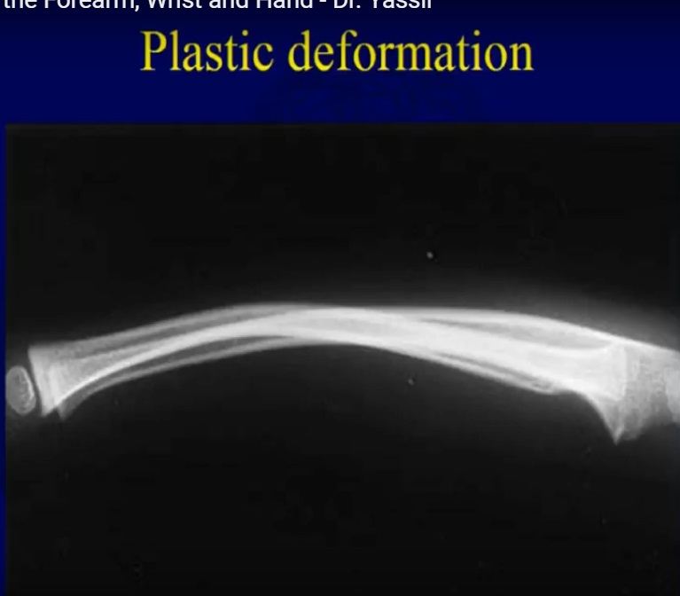
Plastic deformation
- Permanent bending without cortical disruption.
- Periosteum remains intact.
- No callus formation.
- May restrict forearm rotation.
- Remodeling is unreliable.
- Correction is indicated for angulation greater than 20 degrees, functional limitation, or age greater than 8 years.
Greenstick fracture
- One cortex is broken while the opposite cortex remains intact.
- Includes buckle (torus) fractures.
- Intact cortex tends to recoil after reduction.
- Usually treated with a well molded long arm cast.
- Completion of the fracture under anesthesia may occasionally be required.
Complete fracture
- Both cortices are disrupted.
- Highly unstable.
- Proximal fragment is usually supinated by the biceps.
- Distal fragment is pronated by the pronator muscles.
Goals of Treatment
- Restore alignment and cosmetic appearance.
- Preserve soft tissues.
- Relieve pain.
- Achieve functional forearm rotation.
- Minimum functional motion is 50 degrees each of pronation and supination.
- Normal motion is approximately 90 degrees each of pronation and supination.
Principles of Closed Reduction
- Apply traction and countertraction.
- Exaggerate the deformity to unlock the fragments.
- Correct rotational deformity.
- Point the thumb toward the apex of the deformity.
- Apex volar deformity requires pronation.
- Apex dorsal deformity requires supination.
Acceptable Alignment
- Avoid angulation greater than 25 degrees whenever possible.
- Acceptable alignment depends on age, fracture location, and remodeling potential.
- Preserve the interosseous space between the radius and ulna.
Remodeling Potential
Factors affecting remodeling
- Younger age.
- Fractures closer to the physis.
- Sagittal plane deformities remodel best.
- Coronal deformities remodel less.
- Rotational deformities do not remodel.
Remodeling rate
- Approximately 1 degree per month.
Follow Up After Reduction
- Weekly radiographs for the first 3 weeks.
- Shaft fractures may be remanipulated up to 3 weeks.
- Distal physeal fractures should be remanipulated only within 7 to 10 days.
Principles of Casting
- Three point molding.
- Cast index should be less than 0.8.
- Maintain a straight ulnar border.
- Maintain an interosseous mold.
Cast saw burns
- Caused by heat transfer through a wet cast.
- More common in sedated children and wet fiberglass casts.
- Prevent by allowing the cast to dry, using a protective strip, and cooling the saw blade frequently.
Complications
- Malunion is the most common complication.
- Refracture.
- Compartment syndrome.
- Radioulnar synostosis.
- Neurovascular injury.
Indications for Surgery
- Open fractures.
- Compartment syndrome.
- Irreducible fractures.
- Loss of reduction.
- Polytrauma or floating elbow.
- Neurovascular compromise.
- Refracture.
Surgical Options
Intramedullary nailing
- Preferred surgical technique.
- Titanium elastic nails are commonly used.
- Causes minimal soft tissue injury.
- Provides good stability.
- Nails should be prebent to approximately three times the bone diameter.
Plate fixation
- Preferred for older children.
- Useful for comminuted fractures.
- Indicated for some open injuries.
- Higher refracture risk after implant removal.
Galeazzi Fracture
Definition
- Radial shaft fracture associated with distal radioulnar joint disruption.
Clinical features
- Prominent distal ulna.
- Distal radioulnar joint instability.
Management
- Reduce the radial fracture.
- Assess distal radioulnar joint stability.
- Fix the distal radioulnar joint if instability persists.
Distal Radius Fractures
Types
- Metaphyseal fractures are the most common.
- Physeal fractures, with Salter Harris Type II being the most common.
- Buckle (torus) fractures.
Management
Non displaced fractures
- Short arm cast for approximately 3 weeks.
- Buckle fractures are treated with a removable splint.
Displaced fractures
- Closed reduction followed by casting.
- Hematoma block is a useful anesthetic technique.
Important Points
- Avoid repeated reductions because of the risk of growth arrest.
- Up to 15 to 20 degrees of angulation may be accepted if substantial growth remains.
- Bayonet apposition is acceptable in young children with good remodeling potential.
Complications
- Growth arrest in approximately 5 percent.
- Malunion is uncommon because of excellent remodeling.
- Ulnar overgrowth may produce positive ulnar variance.
Exam Pearls
- Distal radius fracture is the most common pediatric fracture.
- Greenstick and buckle fractures are the most common fracture patterns.
- Remodeling is best in younger children, near the physis, and in the sagittal plane.
- Rotational deformity does not remodel.
- Bayonet apposition is often acceptable in younger children.
- Avoid repeated reductions of physeal fractures to minimize the risk of growth arrest.
Leave a Reply