• Skip to main content
  • Skip to secondary menu
  • Skip to primary sidebar
OrthopaedicPrinciples.com

OrthopaedicPrinciples.com

Integrating Principles and Evidence

Integrating Principles and Evidence

  • Home
  • Editorial Board
  • Our Books
    • Evidence Based Orthopaedic Principles
  • Courses
  • Exams
  • Reviews
  • Live Program
  • Contact

Osteoclast and its Functions

Courtesy; Prof Nabil Ebraheim, University of Toledo, Ohio, USA

The bone has two important cells – osteoclast and osteoblasts.


  • Osteoclasts are irregular shaped giant cells that break down or reabsorbs bone.
  • Osteoblasts are cells that are responsible for new bone formation.
  • Osteoclast arise from either macrophages or monocytes .
  • Monocytes fuse together to form multinucleated osteoclast cells.
  • Osteoclast cells have a ruffled border which touches the bone and increases the surface area for absorption of bone.
  • Osteoclast bind to the bone via integrin [protein].
  • Vitronectin helps to attach the osteoclasts to the bone.
  • The bone is absorbed at the howship lacunae .
  • As the ruffled border of the osteoclast contacts the bone it secretes acids that lowers the pH level and the osteoclast then absorbs the mineralized bone matrix.
  • Cathepsin K is an enzyme that removes the bone at the ruffled border.
  • Osteoclast secrete tartate resistant acid phosphates.
  • Osteoclasts are unable to absorb unmineralized osteoid.
  • There are proteins that interact with the osteoclasts and osteoblasts to control bone reabsorption.
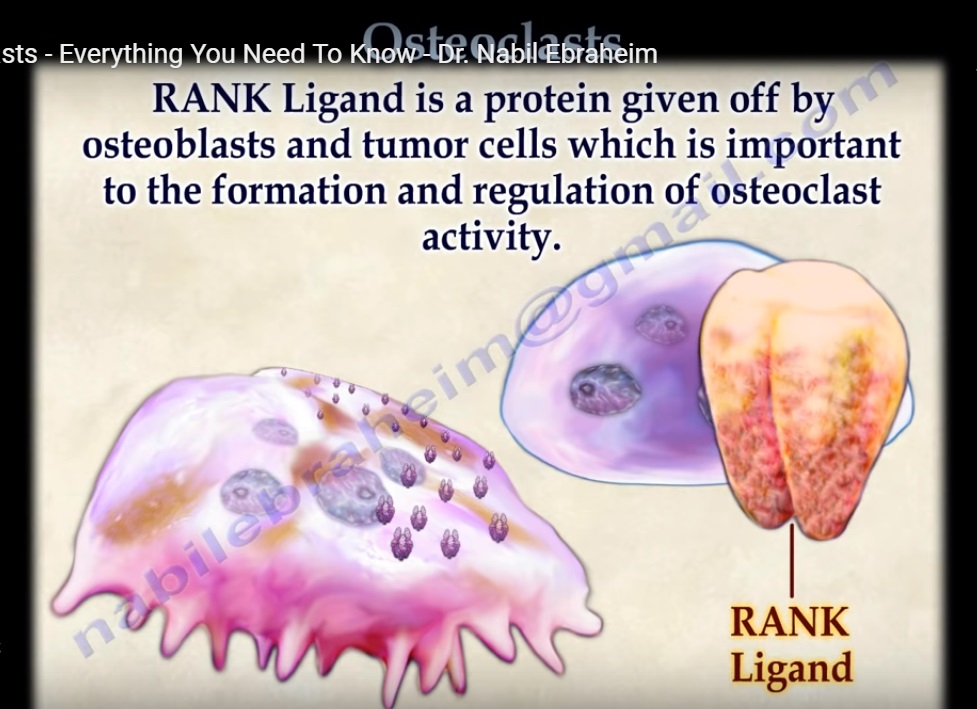
  • The osteoclast has RANK receptors on its surface.
  • RANK ligand is a protein given off by the osteoblasts and tumor cells which is important to the formation and regulation of osteoclast activity.
  • RANK ligand proteins interact with the RANK receptors on the surface of the osteoclast.
  • OPG is another protein that is secreted by the osteoblasts that block the binding of RANK ligand to RANK..
  • The OPG protein is a decoy that stops the osteoclast from differentiation, fusion and activation , which causes decrease in bone reabsorption and destruction. This action of blocking the binding of RANK ligand to RANK reduces osteoclast activity. Excessive amounts of RANK ligand leads to more osteoclasts and greater bone loss.

Osteoclast inhibition

  • Calcitonin- there is a receptor for calcitonin on the osteoclast surface that inhibits bone resorption.
  • Bisphosphonates- prevents formation of the ruffled border and may cause apoptosis
  • IL- 10- suppress osteoclast activity.
  • OPG- blocks the binding of RANK ligand to RANK.
  • Transforming growth factor beta– inceases OPG
  • Estrogen- decreases RANK ligand expression by the osteoclast. decreases activity of the adenylyl cyclase

Osteoclast activation

  • Parathyroid hormone related protein
  • Secreted by many cancer cells including breast cancer. Utilizes adenyl cyclase. Bind to a receptor of the osteoblast to produce RANK ligand.
  • 1,25 dihydroxy vit D- increases production of RANK ligand
  • IL_6-tumors especially myeloma
  • Prostaglandin E2-activate adenylyl cyclase
Post Views: 6,583

Related Posts

  • Osteoclast function and Roles

    ? Courtesy: Prof Nabil Ebraheim, University of Toledo, Ohio, USA Osteoclast is a large multinucleated…

  • Sciatica Anatomy

    Courtesy: Prof Nabil Ebraheim University of Toledo, Ohio, USA Anatomy of sciatica The sciatic nerve…

  • Acetabular fractures: Applied Anatomy

    Courtesy: Saqib Rehman MD Director of Orthopaedic Trauma Temple University Philadelphia Pennsylvania USA

Reader Interactions

Comments

  1. kiran patel says

    at

    GOOD

Leave a Reply

Your email address will not be published. Required fields are marked *

Primary Sidebar

Follow Us

instagram slideshare

Categories

  • -Applied Anatomy
  • -Approaches
  • -Basic Sciences
  • -Cartilage & Meniscus
  • -Classifications
  • -Examination
  • -Foot and Ankle
  • -Foot and Ankle Trauma
  • -FRCS(Tr and Orth) tutorials
  • -Gait
  • -Hand and Wrist
  • -Hand and Wrist Trauma
  • -Hand Infections
  • -Hip and Knee
  • -Hip Preservation
  • -Infections
  • -Joint Reconstruction
  • -Knee Arthroplasty
  • -Knee Preservation
  • -Metabolic Disorders
  • -Oncology
  • -OrthoBiologics
  • -OrthoPlastic
  • -Paediatric Orthopaedics
  • -Paediatric Trauma
  • -Patellofemoral Joint
  • -Pelvis
  • -Peripheral Nerves
  • -Principles
  • -Principles of Surgery
  • -Radiology
  • -Rheumatology
  • -Shoulder and Elbow
  • -Shoulder and Elbow Arthroplasty
  • -Spine Deformity
  • -Spine Oncology
  • -Spine Trauma
  • -Spine, Pelvis & Neurology
  • -Sports Ankle and Foot
  • -Sports Elbow
  • -Sports Knee
  • -Sports Medicine
  • -Sports Medicine Hip
  • -Sports Shoulder
  • -Sports Wrist
  • -Statistics
  • -Technical Tip
  • -Technology in Orth
  • -Trauma
  • -Trauma (Upper Limb)
  • -Trauma Life Support
  • -Trauma Reconstruction
  • Book Shelf
  • Book Shelf Medical
  • Careers
  • Case Studies and Free Papers
  • DNB Ortho
  • Evidence Based Orthopaedic Principles
  • Evidence Based Orthopaedics
  • Exam Corner
  • Fellowships
  • Guest Editor
  • Guest Reviews
  • Image Quiz
  • Instructional Course Lectures
  • Journal Club
  • MCQs
  • Meetings and Courses
  • Multimedia
  • News and Blog
  • Plaster Techniques
  • Podcasts
  • Public Health
  • Rehabilitation
  • Research
  • Shorts and Reels
Copyright@orthopaedicprinciples.com. All right rerserved.