Courtesy: Prof Nabil Ebraheim, University of Toledo, Ohio, USA
Osgood-Schlatter Disease
Introduction
Osgood-Schlatter disease is a common overuse condition in adolescents characterized by:
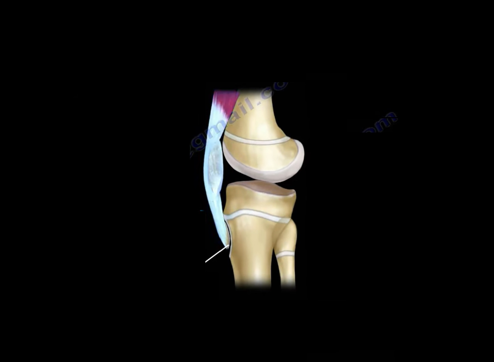
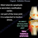
- Traction apophysitis of the tibial tubercle
The condition occurs due to repetitive stress at the insertion of the:
- Patellar tendon
on the developing tibial tubercle.
It is typically seen in active children involved in sports.
Anatomy and Pathophysiology
Tibial Tubercle Anatomy
The tibial tubercle is an:
- Apophysis
which serves as the attachment site for the:
- Patellar tendon
It represents a secondary ossification center.
Mechanism of Injury
Repetitive traction forces from the quadriceps mechanism lead to:
- Inflammation
- Microtrauma
- Pain at the tibial tubercle
This process is aggravated by repetitive activities involving:
- Running
- Jumping
- Sprinting
Ossification Timeline of the Tibial Tubercle
Understanding the developmental stages is important.
Younger Than 11 Years
- Tibial tubercle remains largely cartilaginous
Between 11–14 Years
- Apophysis develops
Between 14–18 Years
- Apophysis gradually fuses with the epiphysis
After 18 Years
- Complete fusion typically occurs
Symptoms usually resolve after skeletal maturity.
Epidemiology
Common Age Groups
Boys
- Commonly affected between 12–15 years
Girls
- Commonly affected between 8–12 years
Girls often present earlier because of earlier skeletal maturation.
Bilateral Involvement
Approximately:
- 20–30% of patients
have bilateral symptoms.
Risk Factors
Osgood-Schlatter disease is commonly associated with sports involving:
- Jumping
- Running
- Soccer
- Sprinting
- Repetitive knee extension
High athletic activity increases traction stress on the tibial tubercle.
Clinical Presentation
Symptoms
Patients commonly report:
- Pain over the tibial tubercle
- Activity-related pain
- Pain worsening during sports
Physical Examination
Typical findings include:
- Swelling over the tibial tubercle
- Local tenderness
- Enlarged tibial tubercle
- Pain with resisted knee extension
Symptoms improve with rest.
Important Clinical Consideration
In cases with:
- Unilateral severe symptoms
- Atypical presentation
other conditions should be excluded, including:
- Infection
- Tumor
- Trauma
Investigations
Plain Radiographs
X-rays may demonstrate:
- Fragmentation of the tibial tubercle
- Irregular ossification
- Soft tissue swelling
Radiographic findings should always be correlated clinically.
Natural History
Osgood-Schlatter disease is generally:
- Self-limiting
Most patients improve after:
- Skeletal maturity
- Closure of the apophysis
Residual prominence of the tibial tubercle may persist.
Management
Conservative Treatment
Non-operative treatment is the mainstay of management.
Activity Modification
Reducing aggravating activities is essential.
Complete cessation of sports is usually unnecessary unless symptoms are severe.
Physiotherapy
Rehabilitation focuses on:
- Quadriceps stretching
- Hamstring stretching
- Strengthening exercises
Improving flexibility reduces traction forces on the tibial tubercle.
Medications
Symptomatic relief may be achieved with:
- NSAIDs
- Ice application
Important Warning: Steroid Injections
Steroid injections should be avoided because they may cause:
- Tendon damage
- Fat necrosis
- Skin atrophy
- Tendon rupture
Surgical Management
Indications
Surgery is rarely required and is considered in approximately:
- 10% of cases
Indications include:
- Persistent symptoms despite conservative treatment
- Skeletal maturity
- Painful ossicle formation
Surgical Procedure
The typical procedure involves:
- Excision of symptomatic ossicles
Surgery is generally reserved for refractory cases.
Differential Diagnosis
Conditions that may mimic Osgood-Schlatter disease include:
- Sinding-Larsen-Johansson Syndrome
- Tibial tubercle fracture
- Infection
- Tumor
- Patellar tendinopathy
Complications
Most patients recover fully, but possible residual issues include:
- Persistent tibial tubercle prominence
- Activity-related discomfort
- Painful ossicles in adulthood
Long-term functional outcomes are usually excellent.
Key Clinical Pearls
- Osgood-Schlatter disease is a traction apophysitis of the tibial tubercle.
- It commonly affects active adolescents.
- Symptoms worsen with running and jumping activities.
- The condition is usually self-limiting.
- Activity modification and physiotherapy are the main treatments.
- Steroid injections should be avoided.
- Surgery is rarely necessary and reserved for persistent symptomatic ossicles after skeletal maturity.
Final Take-Home Message
Osgood-Schlatter disease is a common overuse injury of adolescence caused by repetitive traction at the patellar tendon insertion on the tibial tubercle.
The condition typically presents with:
- Activity-related anterior knee pain
- Tibial tubercle tenderness and swelling
Most patients improve with conservative treatment and gradual skeletal maturation.
Early recognition, reassurance, activity modification, and rehabilitation are the key components of successful

Very good video