• Skip to main content
  • Skip to secondary menu
  • Skip to primary sidebar
OrthopaedicPrinciples.com

OrthopaedicPrinciples.com

Integrating Principles and Evidence

Integrating Principles and Evidence

  • Home
  • Editorial Board
  • Our Books
    • Evidence Based Orthopaedic Principles
  • Courses
  • Exams
  • Reviews
  • Live Program
  • Contact

OATs Procedure for an OCD Lesion of the Capitellum

Courtesy: Dan Zlotolow, Shirner’s hospital for Children, USA

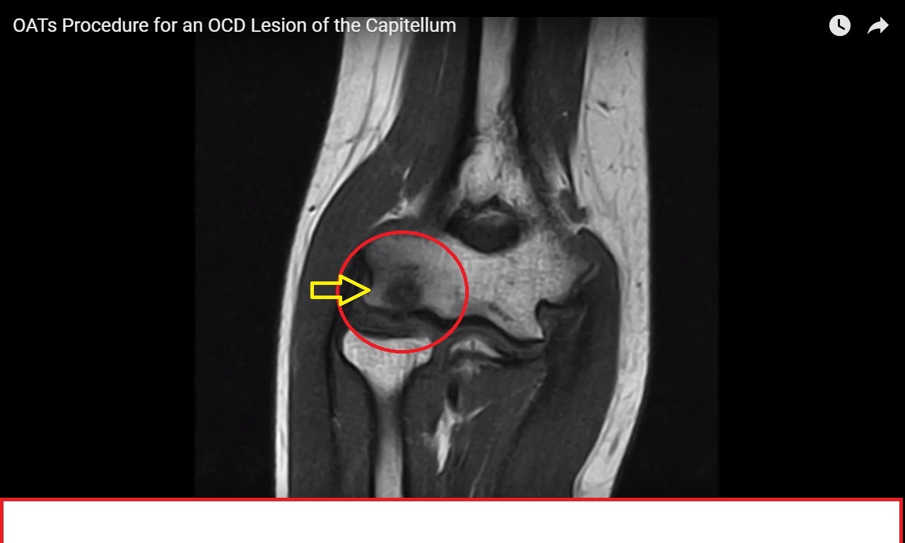
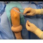
Repetitive load to the outside of the elbow, common in pitchers, gymnast, and wrestlers, can lead to a breakdown of the blood supply to the capitellum of the elbow joint. The capitellum is the end of the humerus bone on the lateral column, articulating with the radial head. The capitellum bears 60% of the weight of the entire elbow joint, and is susceptible to injury from valgus overload. Over time, the loss of blood supply results in avascular necrosis (death) of a portion of the capitellum, leading to pain and loss of motion at the elbow. The area of necrosis is called an osteochondritis dissecans (OCD) lesion. Multiple procedures can be performed to manage OCD lesions, depending on the size and severity. If minimal bone has been lost and the cartilage is damaged, microfracture has shown some good but inconsistent results. If there is a large bony and cartilage defect, these can be replaced with cartilage and bone from the knee, known as an osteochondral autograft technique (OAT). We and others have demonstrated that the OAT procedure has an approximately 90% success rate in returning patients to competitive sports.

Post Views: 7,413

Related Posts

  • Diagnostic Elbow Arthroscopy for OCD lesion of the Capitellum

    Courtesy: Scott Kozin, Dan Zlotolow, Shirner’s hospital for Children, USA This video demonstrates an elbow…

  • Diagnostic Elbow Arthroscopy for OCD lesion of the Capitellum

    Courtesy: Scott Kozin, Dan Zlotolow, Shirner’s hospital for Children, USA This video demonstrates an elbow…

  • Diagnostic Elbow Arthroscopy for OCD lesion of the Capitellum

    Courtesy: Scott Kozin, Dan Zlotolow, Shirner’s hospital for Children, USA This video demonstrates an elbow…

Reader Interactions

Leave a Reply

Your email address will not be published. Required fields are marked *

Primary Sidebar

Follow Us

instagram slideshare

Categories

  • -Applied Anatomy
  • -Approaches
  • -Basic Sciences
  • -Cartilage & Meniscus
  • -Classifications
  • -Examination
  • -Foot and Ankle
  • -Foot and Ankle Trauma
  • -FRCS(Tr and Orth) tutorials
  • -Gait
  • -Hand and Wrist
  • -Hand and Wrist Trauma
  • -Hand Infections
  • -Hip and Knee
  • -Hip Preservation
  • -Infections
  • -Joint Reconstruction
  • -Knee Arthroplasty
  • -Knee Preservation
  • -Metabolic Disorders
  • -Oncology
  • -OrthoBiologics
  • -OrthoPlastic
  • -Paediatric Orthopaedics
  • -Paediatric Trauma
  • -Patellofemoral Joint
  • -Pelvis
  • -Peripheral Nerves
  • -Principles
  • -Principles of Surgery
  • -Radiology
  • -Rheumatology
  • -Shoulder and Elbow
  • -Shoulder and Elbow Arthroplasty
  • -Spine Deformity
  • -Spine Oncology
  • -Spine Trauma
  • -Spine, Pelvis & Neurology
  • -Sports Ankle and Foot
  • -Sports Elbow
  • -Sports Knee
  • -Sports Medicine
  • -Sports Medicine Hip
  • -Sports Shoulder
  • -Sports Wrist
  • -Statistics
  • -Technical Tip
  • -Technology in Orth
  • -Trauma
  • -Trauma (Upper Limb)
  • -Trauma Life Support
  • -Trauma Reconstruction
  • Book Shelf
  • Book Shelf Medical
  • Careers
  • Case Studies and Free Papers
  • DNB Ortho
  • Evidence Based Orthopaedic Principles
  • Evidence Based Orthopaedics
  • Exam Corner
  • Fellowships
  • Guest Editor
  • Guest Reviews
  • Image Quiz
  • Instructional Course Lectures
  • Journal Club
  • MCQs
  • Meetings and Courses
  • Multimedia
  • News and Blog
  • Plaster Techniques
  • Podcasts
  • Public Health
  • Rehabilitation
  • Research
  • Shorts and Reels
Copyright@orthopaedicprinciples.com. All right rerserved.