Courtesy: Scott Kozin, Dan Zlotolow, Shirner’s hospital for Children, USA
Diagnostic Elbow Arthroscopy for Capitellar OCD Lesion
Overview
- Diagnostic arthroscopy performed for:
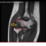
- Osteochondritis Dissecans of Capitellum
- Common in:
- Young patients
- Purpose:
- Assess cartilage integrity
- Identify loose bodies
- Evaluate lesion stability
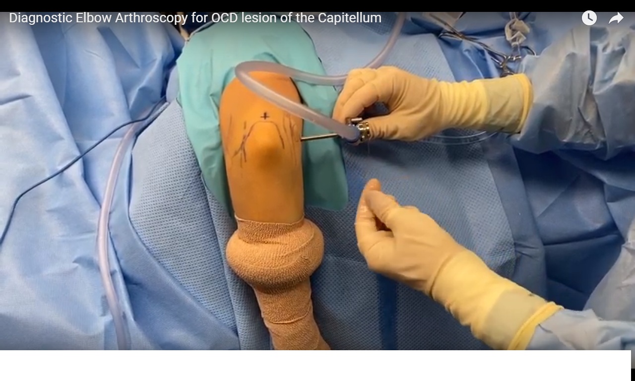
Patient Positioning
- Position:
- Lateral decubitus
Landmarks Identified and Marked
- Ulnar nerve
- Medial epicondyle
- Lateral epicondyle
- Olecranon
Portal Planning
Marked Portals
- Direct posterior portal
- Proximal anteromedial portal
- Posterolateral portal
- Soft spot portal
Additional Step
- Line drawn between:
- Posterolateral portal
- Soft spot portal
Purpose
- Identifies:
- Anconeus muscle location
Preoperative Preparation
- Position and monitor orientation checked
- Limb prepared and draped
- Lower limb prepared (if graft needed)
- Tourniquet applied and inflated
Joint Insufflation
- Fluid injected into joint
Purpose
- Distends capsule
- Improves visualization
Portal Creation and Entry
- Arthroscope introduced via:
- Anteromedial portal
Technique
- Blunt dissection to locate capsule
- Trocar and cannula inserted
- Entry confirmed by:
- Joint fluid outflow
Initial Joint Inspection
Structures Assessed
- Capitellum
- Radial head
- Anterior capsule
- Proximal radioulnar joint
- Trochlea
- Coronoid process
Finding
- No loose bodies detected
Portal Switching Technique
Steps
- Camera advanced across joint
- Switching stick used
- Cannulas inserted over stick
Purpose
- Safe portal exchange
- Minimizes:
- Nerve injury
- Vascular injury
Medial and Lateral Compartment Evaluation
- Trochlea assessed
- Coronoid evaluated
Finding
- No unstable lesions
- No loose bodies
Posterior Compartment Access
Technique
- Posterior portal established
- Care taken to protect:
- Ulnar nerve
Procedure
- Olecranon fossa debrided
- Probe used for assessment
Gutter Evaluation
- Ulnar gutter examined
- Continued to radial gutter
Joint Assessed
- Radiocapitellar joint
Lesion Assessment
Access
- Soft spot portal created
Evaluation Tool
- Probe
Findings
- Lesion:
- Largely healed
- Stable
- Firm
- Non-displaceable
Final Steps
- No surgical intervention required
- Instruments removed
- Portals closed
Key Clinical Points
- Arthroscopy allows:
- Direct visualization
- Assessment of lesion stability
- Stable OCD lesions:
- May not require surgical treatment
- Careful portal placement:
- Reduces neurovascular risk
Final Message
- Diagnostic elbow arthroscopy is a valuable tool for evaluating capitellar OCD lesions and guiding treatment decisions
Leave a Reply