Courtesy: Amr Abdelgawad, Maimonaides Medical Centre, Brooklyn, New York, USA
Femoral Nerve (L2–L4)
- Origin: Lumbar plexus roots L2, L3, L4.
- Motor supply: Quadriceps femoris, iliacus, sartorius, and part of pectineus.
- Primary function: Knee extension and hip flexion.
- Located lateral to the femoral sheath (which contains the femoral artery and vein).
- Travels within the iliopsoas muscle before entering the thigh.
- Compression may occur due to bleeding within the iliopsoas (e.g., hemophilia).
- Differentiating femoral nerve palsy from L3 radiculopathy: test hip adductors (obturator nerve).
Sciatic Nerve (L4–S3)
- Largest nerve in the body.
- Origin: L4, L5, S1, S2, S3 nerve roots.
- Exits pelvis through the greater sciatic foramen below the piriformis muscle.
- Course: deep to piriformis, posterior to external rotators, superficial to quadratus femoris.
- Runs deep to the long head of biceps femoris in the thigh.
- Divides into tibial nerve and common peroneal nerve.
Lateral Femoral Cutaneous Nerve
- Pure sensory nerve from the lumbar plexus.
- Passes under the inguinal ligament.
- Runs between sartorius and tensor fasciae latae.
- Pierces fascia lata to become cutaneous.
- At risk during anterior hip approaches.
- Compression causes meralgia paresthetica.
Tibial Nerve
- Terminal branch of the sciatic nerve.
- Supplies all posterior thigh muscles except the short head of biceps femoris.
- In the leg it supplies posterior compartment muscles: gastrocnemius, soleus, tibialis posterior, flexor digitorum longus, flexor hallucis longus.
- Divides into medial and lateral plantar nerves in the foot.
- Responsible for plantarflexion and toe flexion.
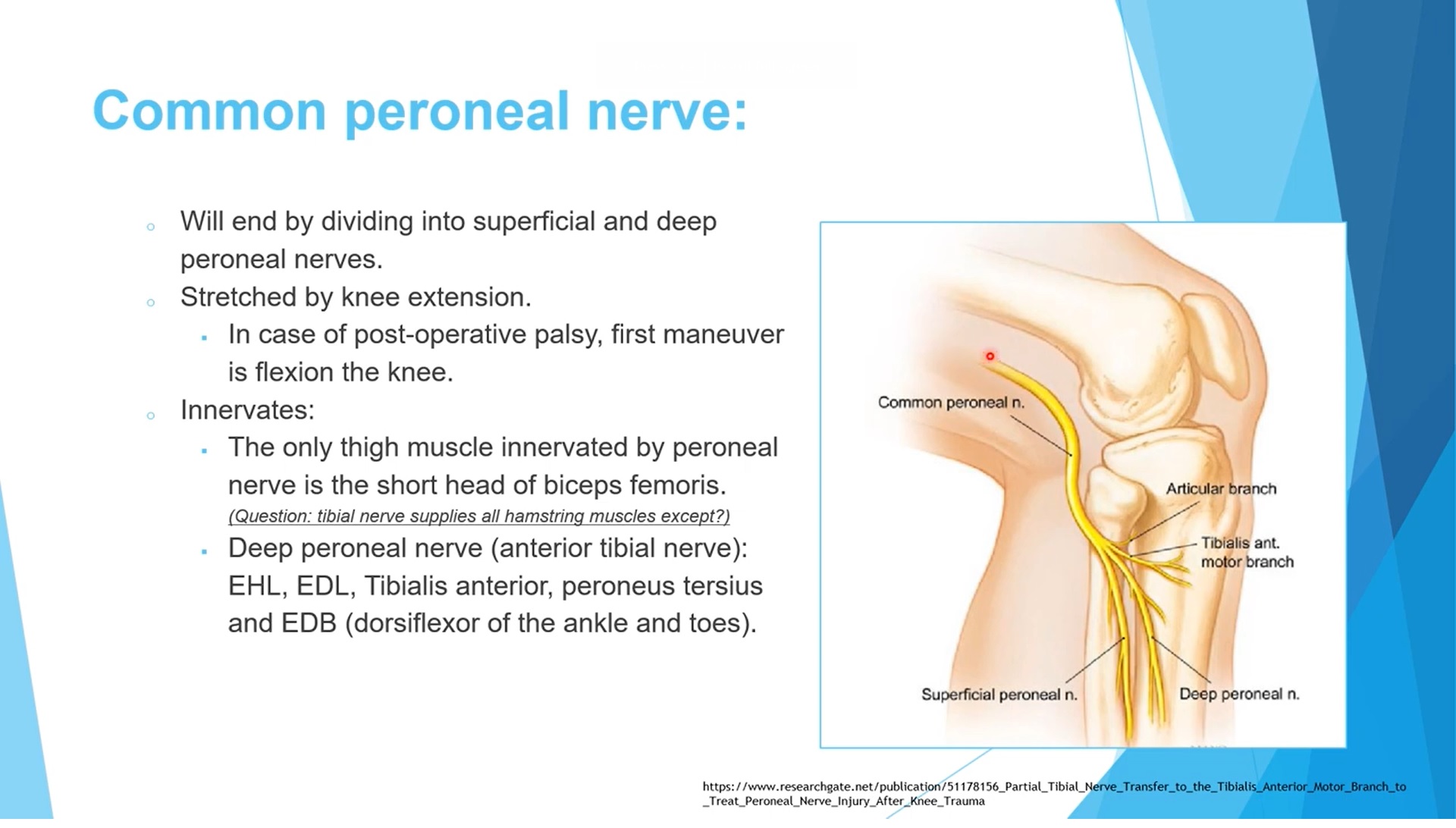
Common Peroneal (Fibular) Nerve
- Branch of the sciatic nerve.
- Supplies the short head of the biceps femoris in the thigh.
- Wraps around the neck of the fibula.
- Divides into deep peroneal and superficial peroneal nerves.
- Highly vulnerable to injury at the fibular neck.
- Stretch increased with knee extension and reduced with knee flexion.
Deep Peroneal Nerve
- Supplies anterior compartment of the leg.
- Muscles: tibialis anterior, extensor hallucis longus, extensor digitorum longus, peroneus tertius.
- Responsible for ankle dorsiflexion and toe extension.
- Provides sensation to the first dorsal web space.
Superficial Peroneal Nerve
- Supplies lateral compartment muscles: peroneus longus and peroneus brevis.
- Responsible for foot eversion.
- Provides sensory innervation to most of the dorsum of the foot.
- Terminates as medial and intermediate dorsal cutaneous nerves.
- Emerges through fascia about 12 cm above the lateral malleolus.
Sural Nerve
- Formed by contributions from the tibial nerve and common peroneal nerve.
- Runs in the posterior calf.
- Provides sensation to the posterolateral leg and dorsolateral foot.
- Small saphenous vein lies medial to the nerve.
- During surgery retracting the vein laterally helps protect the nerve.
Baxter’s Nerve
- Also called the first branch of the lateral plantar nerve.
- Supplies the abductor digiti minimi muscle.
- Can be compressed beneath the abductor hallucis muscle.
- Entrapment causes medial heel pain.
- Often misdiagnosed as plantar fasciitis.
- Tinel’s sign may be present over the nerve.
Saphenous Nerve
- Largest cutaneous branch of the femoral nerve.
- Travels in the adductor canal.
- Infrapatellar branch supplies anterior knee skin.
- Sartorial branch becomes superficial between sartorius and gracilis.
- Commonly injured during medial knee surgeries such as ACL graft harvesting.
Important Root-Level Functions
- L4: tibialis anterior (ankle dorsiflexion) and patellar reflex.
- L5: extensor hallucis longus and gluteus medius.
- S1: gastrocnemius, soleus, and lateral compartment muscles; Achilles reflex.
- S2: perianal sensation.
Dermatomal Sensation of the Foot
- L4: medial foot and great toe.
- L5: dorsum of foot and toes 2–4.
- S1: lateral foot and little toe.
Reflexes
- Patellar tendon reflex: L4.
- Achilles tendon reflex: S1.
- No reliable reflex for L5.
Leave a Reply