
Courtesy: Prof Nabil Ebraheim, University of Toledo, Ohio, USA
Topic / Overview
Comprehensive clinical and anatomical evaluation of peripheral nerve and nerve root injuries around the pelvis and hip joint. The scope addresses mechanisms of injury, localizing neuroanatomy, diagnostic distinctions, and clinical manifestations ranging from Trendelenburg gait to foot drop, meralgia paresthetica, and cauda equina syndrome.
Background / Problems
-
Diagnostic Confounders in Posterior Hip Pain: Posterior hip pain requires differentiation from pathology in adjacent structures, notably the sacroiliac (SI) joint and the lumbar spine.
-
Lumbar Spinal Stenosis: Physical examination frequently reveals minimal objective neurological findings. It is critical to evaluate peripheral pulses to differentiate neurogenic claudication from vascular claudication.
-
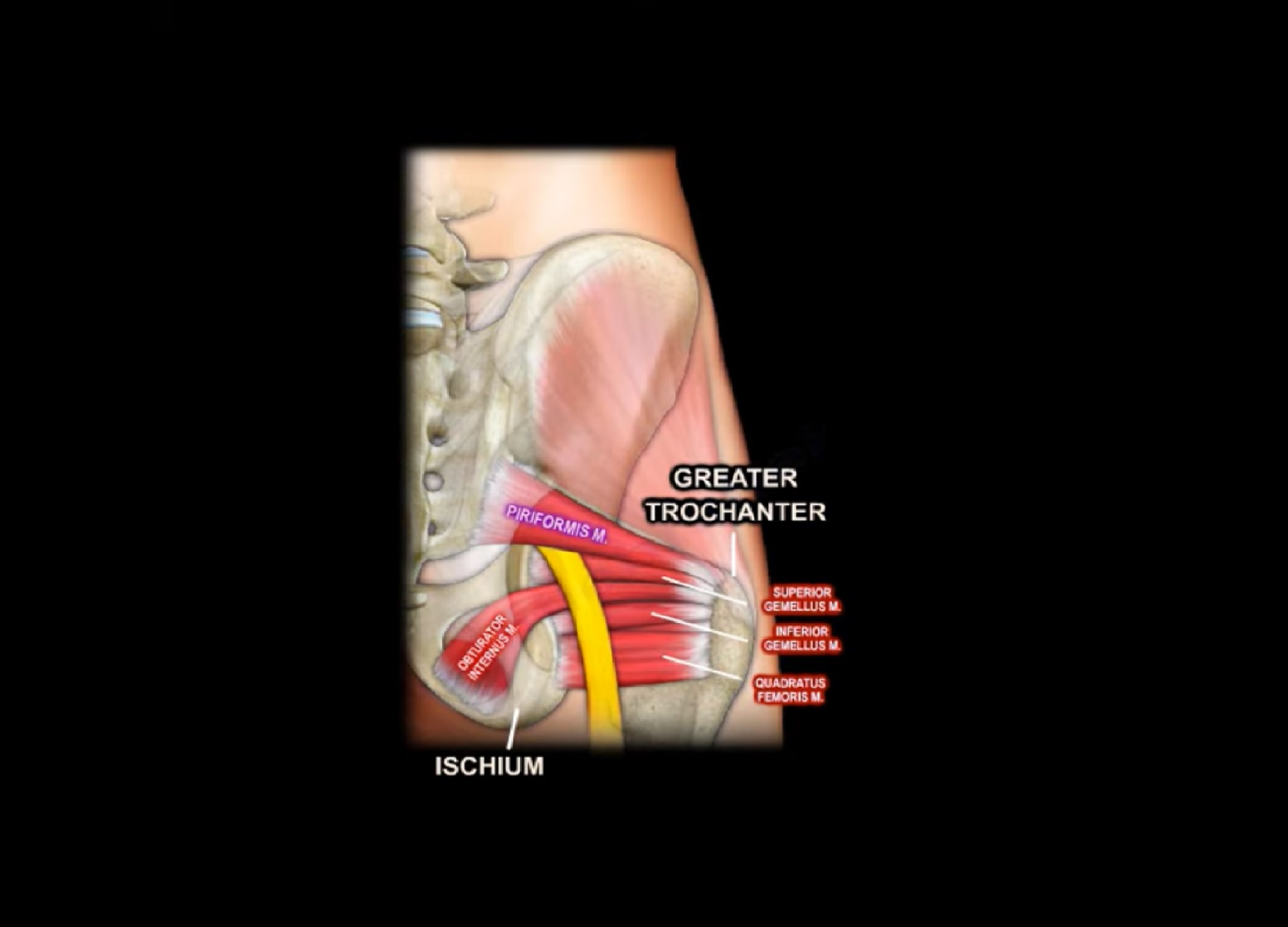
Piriformis Syndrome: Caused by irritation of the sciatic nerve as it passes adjacent to the piriformis muscle, manifesting as posterior hip pain.
-
Sciatica Presentation: A clinical diagnosis characterized by sharp, shooting, electric-shock-like pain radiating down the leg into the foot, accompanied by tingling and numbness. Symptoms are exacerbated by sitting because this position places the nerve on stretch.
Classification and Neuroanatomical Concepts
1. Superior and Inferior Gluteal Nerves
-
Anatomical Boundary: The piriformis muscle acts as the anatomical landmark separating the superior gluteal nerve from the inferior gluteal nerve.
-
Superior Gluteal Nerve:
-
Root Origin: L5 nerve root.
-
Innervation: Hip abductor musculature (and tensor fasciae latae).
-
Pathology: Denervation or muscular failure produces a Trendelenburg gait.
-
-
Inferior Gluteal Nerve:
-
Innervation: Gluteus maximus muscle.
-
2. The L5 Nerve Root Distribution
The L5 nerve root can be compressed by two distinct spinal disc configurations:
-
Ipsilateral Posterolateral Disc Herniation at the L4–L5 intervertebral level.
-
Far Lateral Disc Herniation at the L5–S1 intervertebral level.
Clinical Manifestations of L5 Compromise:
-
Abductor dysfunction and Trendelenburg gait (via superior gluteal innervation).
-
Weakness of great toe extension (extensor hallucis longus).
-
Decreased sensation over the dorsum of the foot (superficial peroneal nerve sensory distribution).
3. Sciatic Nerve and Terminal Branches
-
Root Origin: L4 through S3.
-
Components: Composed of two distinct divisions—the common peroneal (fibular) nerve and the tibial nerve.
-
Common Peroneal Nerve: The division most susceptible to injury along the entire sciatic trunk. Injury manifests clinically as a foot drop.
-
Etiologies of Injury:
-
Pelvic fractures.
-
Lumbosacral plexus injuries.
-
Posterior hip dislocations.
-
4. Anterior Hip Approaches and Sensory Nerves
-
Smith-Petersen Approach: Intermuscular and internervous interval:
-
Interval: Between the tensor fasciae latae (supplied by the superior gluteal nerve) and the Sartorius (supplied by the femoral nerve).
-
Vulnerable Structure: The lateral cutaneous nerve of the thigh (roots L2–L3) runs intimately close to this interval between the sartorius and tensor fasciae latae.
-
-
Meralgia Paresthetica: Entrapment or compression of the lateral cutaneous nerve of the thigh resulting in burning sensation, numbness, or hypesthesia over the anterolateral aspect of the thigh.
5. Femoral Nerve
-
Root Origin: L2, L3, and L4.
-
Terminal Branch: The saphenous nerve is the longest branch of the femoral nerve.
-
Motor Deficit: Denervation leads to quadriceps muscle weakness, leaving the patient unable to actively extend/straighten the knee.
6. Sacral Fractures and Neurologic Deficit Rates
Neurological deficit correlates directly with the anatomical zone of the sacral fracture:
-
Sacral Ala Fractures: Involve the L5 nerve root in approximately 5% of cases.
-
Sacral Foraminal Fractures: Involve sacral nerve roots in approximately 20% to 30% of cases.
-
Sacral Canal Fractures: Produce the greatest neurological deficit; causes cauda equina syndrome with multiple sacral nerve root involvement, presenting with bilateral bowel and bladder dysfunction.
Indications and Patient Selection
Posterior Hip Dislocation
-
Urgency: Demands urgent closed reduction within a strict time threshold of less than 6 hours.
-
Dual Clinical Objectives:
-
Relieve acute mechanical traction/compression on the sciatic nerve.
-
Minimize the risk of avascular necrosis (AVN) of the femoral head.
-
-
Mandatory Action: Perform and document complete neurovascular examinations before and immediately following joint reduction.
Gluteal Abductor Muscle Reconstruction
-
Indication: Irreparable injury or structural deficiency of the hip abductor musculature resulting in chronic abductor insufficiency and a Trendelenburg gait.
-
Surgical Strategy: Muscle transfer of the gluteus maximus (innervated by the inferior gluteal nerve) to reconstruct abductor function.
Technique and Principles
Piriformis Syndrome Management
-
First-Line (Conservative):
-
Physical therapy and targeted muscle stretching protocols.
-
Anti-inflammatory medications.
-
-
Second-Line (Injections):
-
Local therapeutic injections targeted specifically to the buttock and posterior thigh zones.
-
-
Third-Line (Operative):
-
Surgical release/intervention reserved strictly as a last resort.
-
Surgical Intervals: Anterior Approach to the Hip
-
Development of the intermuscular interval for the Smith-Petersen approach requires identification and protection of the lateral cutaneous nerve of the thigh as it courses between sartorius and tensor fasciae latae, respecting the functional boundary between the femoral and superior gluteal nerve territories.
Complications and Limitations
-
Permanent Sciatic Neuropathy / Foot Drop: Protracted traction from an unreduced posterior hip dislocation can result in irreversible common peroneal nerve palsy.
-
Avascular Necrosis: Delays in closed reduction of the hip beyond the 6-hour threshold dramatically elevate the risk of osteonecrosis of the femoral head.
-
Sphincter Incontinence: Sacral canal involvement in high-energy sacral fractures risks permanent loss of bowel and bladder control if cauda equina compression is unaddressed.
-
Iatrogenic Nerve Injury: Suboptimal placement or aggressive retraction along the medial border of the tensor fasciae latae during anterior hip approaches damages the lateral cutaneous nerve of the thigh, yielding painful meralgia paresthetica.
Practical Clinical Implications
-
In patients presenting with a Trendelenburg gait, do not limit evaluation to the hip; systematically assess the L5 nerve root (extensor hallucis longus strength and dorsal foot sensation) and rule out L4–L5 posterolateral or L5–S1 far lateral disc herniations.
-
In suspected lumbar stenosis, always palpate peripheral pulses (dorsalis pedis and posterior tibial) to exclude concurrent vascular claudication.
-
Any patient presenting with high-energy pelvic trauma and a sacral fracture—particularly involving the central sacral canal—must be evaluated for saddle sensory deficit and acute bowel/bladder retention.
Key Take-Home Points
-
Trendelenburg Gait Etiology: Injury to the superior gluteal nerve, hip abductor muscle rupture, or compression of the L5 nerve root (via L4–L5 posterolateral or L5–S1 far lateral disc herniations).
-
Salvage for Abductor Deficiency: Gluteus maximus muscle transfer (utilizing its inferior gluteal nerve supply).
-
Sciatic Nerve Anatomy: Arises from L4–S3; the common peroneal division is the most susceptible component to injury, presenting with foot drop.
-
Hip Dislocation Window: Closed reduction must be achieved in less than 6 hours to avoid femoral head avascular necrosis and permanent sciatic palsy.
-
Sacral Fracture Risk Stratification:
-
Ala: ~5% risk of L5 root injury.
-
Foramina: 20%–30% risk of sacral root deficits.
-
Canal: High risk of cauda equina syndrome with loss of sphincter tone.
-
-
Smith-Petersen Interval: Developed between tensor fasciae latae (superior gluteal nerve) and sartorius (femoral nerve); protect the lateral cutaneous nerve of the thigh (L2–L3) to prevent meralgia paresthetica.

Leave a Reply