
Courtesy: Prof Nabil Ebraheim, University of Toledo, Ohio, USA
L5 Nerve Root Examination
Overview
Examination of any nerve root has three components:
Sensory examination – Motor examination – Reflex examination
For L5, :
- Sensory: dorsum/top of the foot
- Motor: great-toe extension, toe extension, hip abduction, medial hamstrings
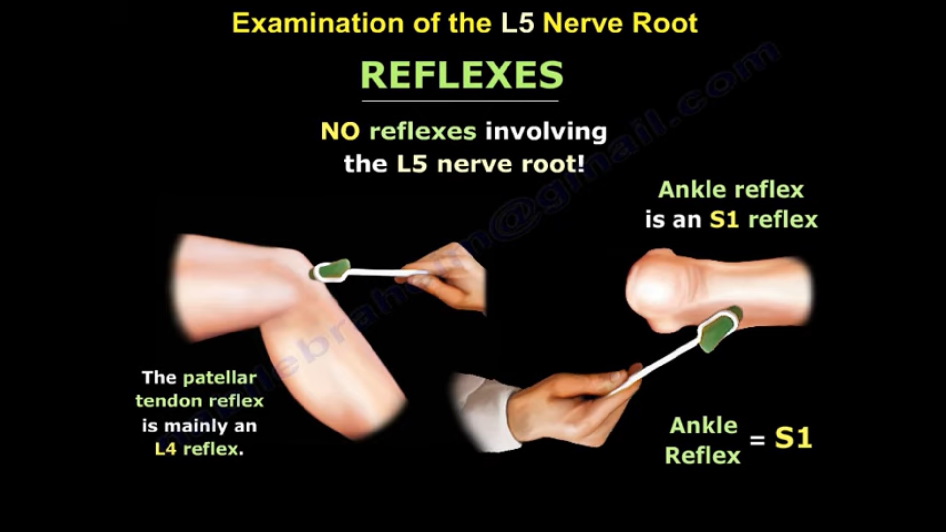
- Reflex: no commonly used specific reflex
- Sensory Examination
gives an easy sensory sequence:
- L4 – medial side of foot
- L5 – dorsum/top of foot
- S1 – lateral side of foot
L5 sensory area
Dorsum of the foot
Quick recall
L4 medial – L5 dorsum – S1 lateral
- Reflex Examination
states:
There is no specific routinely used reflex for L5.
For comparison:
- L4 – patellar reflex
- S1 – Achilles reflex
So in L5 radiculopathy, diagnosis depends more heavily on:
- Sensory examination
- Motor examination
- Provocative tests
- Motor Examination
lists several muscles with important L5 contribution.
Distal muscles
- Extensor hallucis longus
- Extensor digitorum longus
- Extensor digitorum brevis
The most useful practical test is:
Great-toe extension
Extensor Hallucis Longus
Function:
Extension of the great toe
Test by asking the patient to extend the great toe against resistance.
Viva Pearl
L5 – EHL – great-toe extension
Extensor Digitorum Longus
Function:
Extension of the lesser toes
Therefore weakness of toe extension may accompany L5 radiculopathy.
- Proximal L5 Muscles:
- Gluteus medius
- Medial hamstrings:
- Semimembranosus
- Semitendinosus
Therefore, L5 radiculopathy may produce:
- Weak hip abduction
- Weak knee flexion
- Weak great-toe extension
- Hip Abduction
The main muscles discussed are:
- Gluteus medius
- Gluteus minimus
They are supplied through the:
Superior gluteal nerve
L5 involvement may weaken hip abduction.
This can produce a:
Trendelenburg gait
- Trendelenburg Gait
explains:
When the patient stands on the affected limb:
- The pelvis drops on the opposite side
- The trunk leans toward the affected side
The trunk lean acts as a compensatory mechanism.
Quick Recall
Stand on weak side – opposite pelvis drops – trunk leans toward weak side
- Hamstring Examination
describes testing the medial hamstrings by allowing the lower limbs to hang freely and asking the patient to:
Flex the knee
Then palpate medially for contraction of:
- Semimembranosus
- Semitendinosus
These are discussed as having important L5 contribution.
contrasts this with the lateral hamstring:
Biceps femoris, which associates more with S1.
- Ankle Dorsiflexion
states that L5 may also contribute to:
Ankle dorsiflexion
However, in framework, dorsiflexion is considered more predominantly an L4 action.
Therefore:
- L4 – tibialis anterior / ankle dorsiflexion
- L5 – especially great-toe extension
This distinction is useful clinically.
- Clinical Features of L5 Radiculopathy
An L5 lesion may cause:
Sensory
- Decreased sensation over the dorsum of foot
Motor
- Weak great-toe extension
- Weak toe extension
- Weak hip abduction
- Weak knee flexion
- Possible ankle dorsiflexion weakness
- Possible foot drop
Gait
- Trendelenburg gait due to hip-abductor weakness
- Foot-drop gait if dorsiflexion weakness is significant
Reflex
- No specific routinely used L5 reflex
- EMG Findings
states that L5 radiculopathy may show fibrillation/sharp-wave activity in L5-supplied muscles from proximal to distal, including:
- Gluteus medius
- Medial hamstrings
- Extensor hallucis longus
- Extensor digitorum longus
- Extensor digitorum brevis
This pattern can support localization to the L5 root.
- L4–L5 Disc Herniation
Posterolateral L4–L5 Disc
states:
Posterolateral L4–L5 disc herniation ? traversing L5 root
This is the classic disc-root relationship emphasized in the lecture.
Expected findings
- Dorsal-foot sensory disturbance
- Weak great-toe extension
- Possible weakness of hip abduction
- Positive SLR
Foraminal L4–L5 Disc
contrasts this with:
Foraminal L4–L5 disc herniation ? exiting L4 root
Therefore:
| Disc pattern | Root affected |
| L4–L5 posterolateral | L5 |
| L4–L5 foraminal/far lateral | L4 |
- L5–S1 Isthmic Spondylolisthesis
L5–S1 isthmic spondylolisthesis may cause L5 radiculopathy.
- Straight Leg Raise
When L5 is irritated by disc herniation, the patient may have:
- Sciatica
- Positive straight-leg raise
associates the SLR tension sign with:
L5 and S1 nerve-root irritation
Positive SLR
describes reproduction of:
- Leg pain
- Paresthesia
at approximately:
30–70° of hip flexion
Exam Pearl
SLR – lower lumbar/sacral radiculopathy, especially L5/S1
- Femoral Stretch Test
contrasts SLR with the:
Femoral nerve stretch test
which is used more for the upper lumbar roots, particularly around L3/L4 in this lecture series.
Therefore:
- SLR – L5/S1
- Femoral stretch – upper lumbar roots
High-Yield L5 Table
| Component | L5 |
| Sensory | Dorsum of foot |
| Main motor test | Great-toe extension |
| Muscle | EHL |
| Other motor | EDL, EDB |
| Proximal motor | Gluteus medius, medial hamstrings |
| Hip movement | Abduction |
| Knee movement | Flexion contribution |
| Reflex | No specific routinely used reflex |
| Gait abnormality | Trendelenburg gait |
| Possible additional deficit | Foot drop |
| Provocative test | Straight-leg raise |
| Common posterolateral disc | L4–L5 |
L4 vs L5 vs S1 Quick Comparison
| Feature | L4 | L5 | S1 |
| Sensation | Medial foot | Dorsum of foot | Lateral/plantar foot |
| Key motor | Ankle dorsiflexion | Great-toe extension | Plantar flexion |
| Proximal motor | Quadriceps | Hip abductors / medial hamstrings | — |
| Reflex | Patellar | None specific | Achilles |
| Provocative test | Femoral stretch | SLR | SLR |
Practical Examination Sequence
SENSORY
Check dorsum of foot
MOTOR — DISTAL
Great-toe extension – EHL
Toe extension – EDL
MOTOR — PROXIMAL
Hip abduction – gluteus medius
Knee flexion – medial hamstrings
REFLEX
No specific L5 reflex
GAIT
Look for Trendelenburg gait and foot drop
PROVOCATIVE
Straight-leg raise
LOCALIZE DISC
Posterolateral L4–L5 – L5
Key Take-Home Points
- L5 root examination consists of sensory + motor + reflex assessment.
- L5 sensory territory = dorsum of the foot.
- There is no commonly used specific L5 reflex.
- The best practical distal motor test is great-toe extension using EHL.
- Other L5 muscles include EDL, EDB, gluteus medius and medial hamstrings.
- L5 weakness can produce Trendelenburg gait through hip-abductor weakness.
- L5 involvement can also contribute to foot drop.
- Posterolateral L4–L5 disc – traversing L5 root.
- Foraminal L4–L5 disc affects the exiting L4 root.
- SLR is useful for L5/S1 root irritation.
Exam Pearls
L5 = dorsum of foot + EHL + no reflex
L5 motor = great-toe extension
L5 proximal clue = hip abduction weakness
Trendelenburg gait can occur in L5 radiculopathy
L4–L5 posterolateral disc – L5
SLR – L5/S1

Leave a Reply