
Courtesy: Dr. Sandeep Patwardhan, Dr Ashok Shyam, Ortho TV
Pediatric Radial Neck Fractures & Post-Traumatic Proximal Radioulnar Synostosis
Pitfalls in Pediatric Radial Neck Fracture Management
-
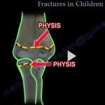
The High Vulnerability Zone: Fractures through the radial neck in skeletally immature patients (ages 7 to 9) carry significant risks of severe complications, specifically proximal radioulnar synostosis (cross-union) and complete loss of forearm pronosupination.
-
The Hazards of Open Reduction:
-
Formal open reduction of a pediatric radial neck fracture significantly increases the risk of periarticular scarring, devascularization of the radial head, and heterotopic cross-union.
-
Direct surgical exposure strips periosteum, introduces surgical hematoma, and is considered a primary contributor to post-traumatic synostosis.
-
-
The Hidden Risk in Closed Management:
-
Non-operative management or closed reduction in a cast is generally preferred over open surgery, but it is not without risk.
-
Concomitant high-energy tear or rupture of the proximal interosseous membrane/capsular structures extending toward the ulna can cause a confluent hematoma that bridges the radius and ulna.
-
Subsequent hematoma ossification can produce a complete radioulnar synostosis even after non-operative treatment in plaster.
-
-
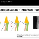
Reduction Strategy: Percutaneous reduction techniques (e.g., Métaizeau intramedullary pin manipulation or percutaneous K-wire joysticking) are favored over open reduction when acceptable closed reduction cannot be achieved, minimizing local periosteal disruption.
Post-Traumatic vs. Congenital Synostosis
-
Congenital Radioulnar Synostosis:
-
Arises from a failure of longitudinal segmentation during embryogenesis.
-
Characterized by primary hypoplasia/absence of the proximal radioulnar joint (PRUJ), abnormal osseous architecture, and contracted soft-tissue envelopes.
-
Simple surgical resection typically fails and has high recurrence rates; rotational osteotomy to place the forearm in a functional position is often preferred.
-
-
Post-Traumatic Radioulnar Synostosis:
-
True secondary osseous bridge formation across a previously normal proximal radioulnar joint.
-
Distinct from congenital forms because articular architecture and soft-tissue envelopes are intact prior to injury.
-
Resection of the synostosis mass can successfully restore active pronosupination when the bone bridge is quiescent and mature.
-
Surgical Protocol: Synostosis Resection & Interposition Arthroplasty
-
Timing of Intervention:
-
Surgical excision is strictly indicated only when the post-traumatic ossification is completely mature and quiescent (cold on biological monitoring, well-corticated on plain radiographs and CT).
-
-
Preoperative Workup & Advanced Imaging:
-
Multiplanar 3D CT imaging is mandatory to map and quantify the exact size, orientation, and longitudinal extent of the fusion mass (e.g., measuring bridge width, typically $\approx 1\text{ cm}$).
-
-
Surgical Approach & Technique:
-
Approach: Posterior/posterolateral exposure to safely access the proximal radius and ulna while protecting the posterior interosseous nerve (PIN).
-
Excision: Isolate the radial head and neck; completely resect the synostosis bridge down to normal cortical margins on both the radius and ulna.
-
Intraoperative Assessment: Confirm immediate, full mechanical restoration of active/passive forearm pronation and supination on the table before reconstruction.
-
-
Interposition Fat Grafting:
-
Excision alone leaves two raw, bleeding cancellous surfaces adjacent to each other, carrying an unacceptably high risk of recurrent cross-union.
-
Harvest an autologous inert fat graft (e.g., from the local triceps area or subcutaneous tissue).
-
Pack and interpose the fat graft securely into the radioulnar interval.
-
Secure the graft with anchoring sutures to prevent migration or washout when releasing the tourniquet.
-
-
Postoperative Rehabilitation:
-
Initiate aggressive, early active and active-assisted range-of-motion physiotherapy within the first few postoperative weeks to prevent recurrent adhesion formation.
-
Classification & Clinical Summary
-
Anatomical Levels: Post-traumatic pediatric cross-unions are classified topographically by location:
-
Proximal (involving the radial neck/bicipital tuberosity and proximal ulna).
-
Diaphyseal (middle third of the forearm).
-
Distal (distal radioulnar joint / metaphyseal region).
-
-
Core Takeaways:
-
Avoid unnecessary open reduction of radial neck fractures in children due to the high risk of cross-union.
-
Differentiate post-traumatic from congenital synostosis; post-traumatic lesions have viable joint potential.
-
Quiescent post-traumatic proximal synostosis can be managed with bridge resection, autologous interposition fat grafting, and early aggressive postoperative mobilization to restore pronosupination.
-

Leave a Reply