Courtesy Dr Pradeep Moonot, Dr Ashok Shyam, Ortho TV
Case Example
A 30 year old female sustained a minor fall from stairs.
Initial diagnosis:
- Midfoot sprain
- Fracture of the base of the fifth metatarsal
Treatment:
- Cast immobilization for 6 weeks
Outcome:
- Persistent pain and deformity
- Re-presented after 3 months
Final diagnosis:
- Missed Lisfranc injury
Why Are Lisfranc Injuries Missed?
Approximately 20 to 40% of Lisfranc injuries are initially missed.
They are commonly mistaken for:
- Midfoot sprain
- Minor foot injury
Reasons for Missed Diagnosis
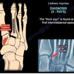
- Subtle radiographic findings
- Polytrauma patients
- Delayed presentation
- Misinterpretation of imaging
- Failure to obtain weight bearing radiographs
Importance of the Lisfranc Joint
The Lisfranc joint complex connects:
- Metatarsal bases
- Cuneiforms
- Cuboid
Consequences of Missed Injury
Untreated Lisfranc injuries may result in:
- Chronic midfoot pain
- Progressive deformity
- Loss of foot function
- Post traumatic arthritis
- Difficulty walking and weight bearing
Factors Influencing Treatment
Management depends on:
- Degree of deformity
- Number of joints involved
- Soft tissue damage
- Cartilage injury
- Ligament disruption
- Presence of arthritis
- Functional demands of the patient
Treatment Options
1. Conservative Management
Indications
- Minimal displacement
- Low energy injury
- Low demand patients
Limitations
- Painful malunion
- Progressive deformity
- Functional impairment
Conservative treatment is generally reserved for carefully selected cases.
2. Open Reduction and Internal Fixation (ORIF)
Challenges in Delayed Cases
When presentation occurs more than 6 weeks after injury:
- Reduction becomes difficult
- Extensive soft tissue dissection may be required
- Cartilage damage may already exist
- Osteopenia may compromise fixation
Evidence
Several studies demonstrate that good outcomes can still be achieved even months after injury if an anatomical reduction is obtained.
3. Arthrodesis (Fusion)
Indications
- Severe cartilage damage
- Post traumatic arthritis
- High energy injuries
- Severe instability
- Failed previous treatment
Advantages
- Eliminates painful arthritic joints
- Provides durable stability
Disadvantages
- Loss of normal joint motion
- Increased stress on adjacent joints
- Potential secondary arthritis
Motion Considerations
Medial Column
- First TMT joint normally has approximately 4 to 6 degrees of motion
- Motion contributes to normal gait
Lateral Column
- Requires greater mobility
- Fusion may lead to stiffness and altered foot mechanics
4. Biological Reconstruction (Ligament Reconstruction)
Principle
Reconstruction of the Lisfranc ligament using tendon grafts such as:
- Gracilis tendon
Indications
- Predominantly ligamentous instability
- Preserved articular cartilage
- Delayed presentation without advanced arthritis
Reported Outcomes
Studies have shown:
- Reduced pain
- Good functional recovery
- Return to sports
- Good outcomes despite delayed surgery
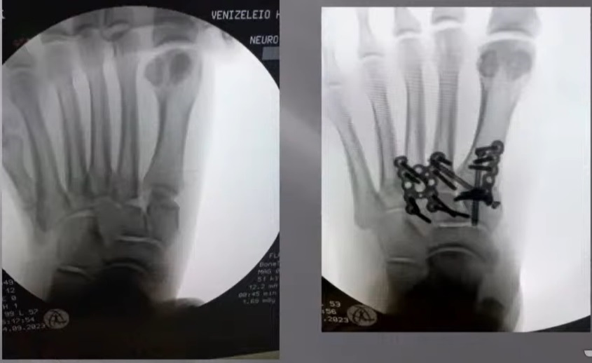
Surgical Example
Findings
- Subtle widening between first and second metatarsals
- Subluxation of the second tarsometatarsal joint
Procedure
- Anatomical reduction
- Medial plate fixation
- Dorsal plate stabilization
Outcome
6 Months
- Stable reduction maintained
1.5 Years
- Alignment preserved
- Pain free foot
- High patient satisfaction
Outcomes of Missed Lisfranc Injuries
Even after treatment:
- Approximately two thirds of patients achieve good results
- One third continue to experience symptoms
This highlights the importance of early diagnosis.
Management Algorithm for Delayed Lisfranc Injury (> 6 Weeks)
Conservative Treatment
Suitable for:
- Low demand patients
- Minimal displacement
- Stable deformity
ORIF ± Ligament Reconstruction
Best suited for:
- Low energy injuries
- Medial column involvement
- Preserved cartilage
- No significant arthritis
- Predominantly ligamentous instability
Arthrodesis
Preferred when:
- High energy trauma
- Severe deformity
- Advanced cartilage damage
- Established arthritis
- Chronic painful instability
Key Clinical Message
- Lisfranc injuries are missed in 20 to 40% of cases.
- Weight bearing radiographs are essential when Lisfranc injury is suspected.
- Delayed diagnosis significantly increases treatment complexity.
- Anatomical reduction remains the most important determinant of outcome.
- Early diagnosis provides the best chance of restoring normal foot function.
Exam Pearls
- Lisfranc injury involves the tarsometatarsal joint complex.
- Up to 40% are initially missed.
- Plantar ecchymosis is an important clinical sign.
- Weight bearing radiographs should be obtained whenever possible.
- Untreated injuries lead to chronic pain, deformity, and arthritis.
- Delayed injuries may require ORIF, ligament reconstruction, or arthrodesis.
- Anatomical reduction is the strongest predictor of a successful outcome.

Leave a Reply