
Courtesy: Prof Nabil Ebraheim, University of Toledo, Ohio, USA
Complications and Management of Femoral Neck Fractures
1. Total Hip Arthroplasty Versus Hemiarthroplasty
Total hip arthroplasty (THA) has an important role in the management of displaced femoral neck fractures in selected elderly patients.
Indications for Total Hip Arthroplasty
THA may be considered in:
- Physiologically active elderly patients.
- Patients with a displaced femoral neck fracture.
- Patients who were independently mobile before the fracture.
- Patients with sufficient physiological reserve to tolerate the procedure.
Compared with hemiarthroplasty, THA may provide:
- Better long-term functional outcomes in appropriately selected patients.
- A lower risk of revision surgery.
However, THA may carry a higher risk of dislocation.
2. Failure of Fixation
The quality of fracture reduction and fixation is critical when treating femoral neck fractures with internal fixation.
Principles of Fixation
- Reduction should be anatomic.
- Closed reduction may initially be attempted.
- If an acceptable anatomical reduction cannot be obtained, open reduction should be performed.
Open reduction can be performed through approaches such as:
- Anterior approach.
- Watson-Jones approach.
Management of Failed Fixation
When fixation fails, options include:
- Revision fixation in appropriately selected patients.
- Prosthetic replacement, particularly in elderly patients.
In elderly patients with displaced femoral neck fractures treated with screw fixation, failure and subsequent revision may be substantial.
3. Fracture Distal to the Fixation
A fracture may occasionally occur distal to the site of femoral neck fixation.
Possible contributing factors include:
- Screws positioned at or below the lesser trochanter.
- Poor bone quality.
- Suboptimal screw trajectory.
- Excessive anterior positioning of screws.
- Multiple drilling or guidewire attempts.
- Local weakening of the femur around the fixation.
Management
Treatment depends on:
- Location and pattern of the new fracture.
- Stability of the original femoral neck fracture.
- Bone quality.
- Patient age and physiological status.
Options may include revision fixation of the femoral neck together with fixation of the associated subtrochanteric or distal fracture.
4. Nonunion of the Femoral Neck
Femoral neck fractures are intracapsular fractures and have a recognized risk of nonunion.
The fracture environment contributes to this risk because:
- The fracture is surrounded by synovial fluid.
- There is limited extraosseous blood supply.
- There is little or no external callus formation.
- Healing therefore depends predominantly on direct/intraosseous bone healing.
Clinical Presentation
Nonunion may present with:
- Persistent or recurrent groin pain.
- Pain with hip extension.
- Pain during weight-bearing.
- Persistent functional limitation.
Incidence
The risk of nonunion is approximately:
- 5% in nondisplaced fractures.
- Up to 25% in displaced fractures, according to the figures presented in the lecture.
5. Management of Femoral Neck Nonunion
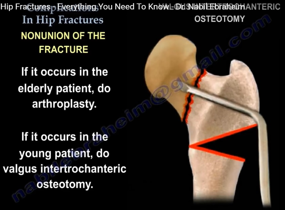
Treatment depends strongly on the patient’s age and the condition of the femoral head.
Elderly Patients
In elderly patients, treatment will frequently involve:
- Hemiarthroplasty, or
- Total hip arthroplasty.
The choice depends on patient factors and the condition of the hip.
Young Patients
Joint-preserving treatment may be considered in younger patients.
Options include:
- Revision fixation.
- Valgus intertrochanteric osteotomy.
- Vascularized fibular grafting in selected cases.
6. Role of Valgus Osteotomy
Nonunion is more common with vertically oriented femoral neck fracture patterns.
A vertical fracture line generates substantial shear forces across the fracture.
A valgus intertrochanteric osteotomy can alter the mechanical environment by:
- Reorienting the fracture line.
- Making the fracture more horizontal.
- Converting shear forces into more favorable compressive forces.
This may facilitate union in appropriately selected young patients.
7. Timing of Diagnosis of Nonunion
Nonunion may become clinically and radiographically apparent during follow-up.
Diagnosis can sometimes be challenging because:
- The femoral neck does not develop prominent periosteal callus.
- Radiographic evidence of union may therefore be less obvious than in extracapsular fractures.
Persistent pain and failure of progressive radiographic healing should raise suspicion for nonunion.
8. Medical Complications
Patients with femoral neck fractures, particularly elderly patients, are at high risk of medical complications.
Venous Thromboembolism
The risk of deep venous thrombosis (DVT) is substantial.
Therefore, thromboprophylaxis should include appropriate:
- Mechanical prophylaxis.
- Pharmacological prophylaxis.
The exact regimen should be based on current guidelines and the individual patient’s bleeding and thromboembolic risk.
9. Multidisciplinary Management
Elderly patients with hip fractures frequently have significant medical comorbidities.
Management should therefore involve:
- Orthopaedic team.
- Medical/geriatric team.
- Anaesthetic team.
- Physiotherapy and rehabilitation services.
The goals are to:
- Optimize medical conditions.
- Avoid unnecessary surgical delay.
- Mobilize the patient as early as possible.
- Reduce complications associated with prolonged immobilization.
10. Early Surgery and Mobilization
Hip fracture in an elderly patient is associated with significant morbidity and mortality.
The lecture emphasizes the importance of avoiding unnecessary surgical delay.
Early surgery facilitates:
- Earlier mobilization.
- Earlier rehabilitation.
- Reduced complications associated with prolonged bed rest.
- Earlier return to functional independence.
The precise timing of surgery should take into account the patient’s medical condition and the need for appropriate optimization.
11. Osteoporosis Management
A hip fracture in an elderly patient is frequently a fragility fracture and should trigger assessment and treatment of underlying osteoporosis.
Management may include:
- Assessment of bone health.
- Calcium and vitamin D optimization where appropriate.
- Anti-osteoporosis medication when indicated.
- Fall-risk assessment.
- Strength and balance training.
The objective is not only to treat the current fracture but also to reduce the risk of subsequent fragility fractures.
12. Osteonecrosis of the Femoral Head
Another important complication following femoral neck fracture is osteonecrosis (avascular necrosis, AVN) of the femoral head.
The risk is related to disruption of the blood supply to the femoral head.
Blood Supply
The medial femoral circumflex artery is the predominant source of blood supply to the adult femoral head.
Displacement of the fracture can disrupt the retinacular vessels and compromise femoral head perfusion.
13. Clinical Presentation of AVN
Patients may develop:
- Groin pain.
- Proximal thigh pain.
- Mechanical hip symptoms.
- Progressive pain with weight-bearing.
AVN may occur despite apparently successful fracture fixation and union.
14. Incidence of AVN
The lecture cites an incidence of approximately:
- 10% following nondisplaced femoral neck fractures.
- 30% following displaced femoral neck fractures.
The risk is influenced by factors related to the initial injury and treatment.
15. Diagnosis of AVN
MRI
MRI is the most sensitive imaging modality for detecting early osteonecrosis.
Radiographs
Plain radiographs may demonstrate AVN when structural changes have developed.
However:
- Early AVN may have normal radiographs.
- Not every patient with AVN develops femoral head collapse.
Therefore, the presence of osteonecrosis alone does not necessarily mean that the patient requires immediate surgical intervention.
16. Femoral Head Collapse
The clinically important event is structural collapse of the femoral head.
Segmental collapse may become apparent:
- As early as 6–9 months after the fracture in some patients.
- More commonly during the second year.
If collapse does not occur during the early follow-up period, the risk progressively decreases.
17. Factors Associated With AVN
The risk of osteonecrosis is influenced by:
- Initial fracture displacement.
- Severity of the initial injury.
- Time to reduction.
- Quality of reduction.
- Vascular disruption associated with the fracture.
An anatomic reduction and appropriate stabilization remain important principles in the management of femoral neck fractures.
18. Management of AVN in Young Patients
Treatment depends on:
- Patient age.
- Symptoms.
- Size of the necrotic segment.
- Location of the lesion.
- Presence or absence of femoral head collapse.
In younger patients with limited femoral head involvement, joint-preserving procedures may be considered.
Possible options include:
- Core decompression.
- Bone grafting.
- Vascularized fibular grafting.
Total hip arthroplasty may be considered when the femoral head involvement is extensive or when collapse and secondary arthritis have developed.
19. Management of AVN in Elderly Patients
In elderly patients with symptomatic femoral head osteonecrosis following femoral neck fracture, arthroplasty is generally the preferred reconstructive option.
Depending on patient characteristics, this may involve:
- Hemiarthroplasty, or
- Total hip arthroplasty.
20. Intra-articular Screw Penetration
A further complication of internal fixation is penetration of the screws into the hip joint.
This can result in:
- Articular cartilage damage.
- Persistent pain.
- Secondary degenerative changes.
- Failure of fixation.
Prevention
Screw position should be carefully assessed intraoperatively using multiple fluoroscopic views.
The screws should:
- Remain within the femoral head.
- Avoid penetrating the articular surface.
- Be positioned close enough to the subchondral bone to provide effective fixation without entering the joint.
21. Screw Position and Fixation Principles
When using multiple cannulated screws:
- Screws should be appropriately positioned within the femoral neck and head.
- Multiple fluoroscopic views should be used to confirm their position.
- The screws should generally be parallel.
- Parallel screws facilitate controlled compression across the fracture.
Screw Threads
The position of the threads relative to the fracture is important.
The threads should cross the fracture site so that compression can occur.
If the threads engage the fracture surfaces inappropriately, they may prevent appropriate compression or cause distraction.
Both partially threaded and fully threaded screws may have applications depending on the fracture pattern and fixation strategy.
22. Femoral Neck Shortening
Shortening is a recognized consequence of femoral neck fracture fixation.
Although union may be achieved, excessive shortening can result in:
- Altered hip biomechanics.
- Abductor weakness.
- Limb-length discrepancy.
- Reduced walking efficiency.
- Poorer functional outcome.
The lecture emphasizes that a healed femoral neck fracture with significant shortening may still be associated with a poor functional outcome.
Key Take-Home Messages
- Anatomic reduction and stable fixation are fundamental in femoral neck fractures treated with internal fixation.
- Failed closed reduction should prompt consideration of open reduction.
- Elderly patients with displaced femoral neck fractures may be better candidates for arthroplasty than screw fixation.
- Femoral neck nonunion is more common in displaced and vertically oriented fractures.
- Valgus intertrochanteric osteotomy can improve the mechanical environment for union in selected young patients.
- DVT prophylaxis and early mobilization are essential components of hip fracture care.
- Hip fracture should trigger assessment and treatment of osteoporosis.
- AVN remains an important complication, particularly after displaced fractures.
- The medial femoral circumflex artery is the principal blood supply to the femoral head.
- MRI is useful for diagnosing early osteonecrosis.
- Femoral head collapse, rather than osteonecrosis alone, is the major determinant of clinical significance.
- Screw penetration into the joint must be avoided through meticulous positioning and fluoroscopic assessment.
- Femoral neck shortening after fixation may adversely affect long-term hip function.
Leave a Reply