
Courtesy: Raul Lins, Recife, Brazil
Greater Trochanteric Pain Syndrome and Gluteal Tendon Tears
Overview
- Greater trochanteric pain syndrome is a common cause of lateral hip pain.
- It represents a group of disorders affecting the peritrochanteric region rather than a single disease.
- Important causes include:
- Gluteus medius tendinopathy or tear.
- Gluteus minimus tendinopathy or tear.
- Trochanteric bursal pathology.
- Iliotibial band-related disorders.
- External snapping hip.
- Myofascial disorders.
- Gluteal tendon tears are therefore only one component of greater trochanteric pain syndrome.
- Correct identification of the underlying pathology is essential because treatment varies according to the cause.
Greater Trochanteric Pain Syndrome Versus Gluteal Tendon Tear
- Greater trochanteric pain syndrome is a clinical syndrome characterized predominantly by lateral hip pain and tenderness around the greater trochanter.
- Gluteus medius or minimus tears represent a specific structural lesion within this broader syndrome.
- A patient may have greater trochanteric pain syndrome without a gluteal tendon tear.
- Conversely, a significant gluteal tendon tear will generally produce symptoms within the greater trochanteric pain syndrome spectrum.
- Therefore, the terms should not be used interchangeably.
Important Clinical Principle
- Persistent lateral hip pain should not automatically be labelled as trochanteric bursitis.
- Bursal inflammation may accompany gluteal tendinopathy, but tendon pathology is an important underlying cause in many patients.
- Repeated treatment directed only at the bursa may fail if the primary pathology is a gluteal tendon disorder.
- A careful history, physical examination and appropriate imaging are required to establish the diagnosis.
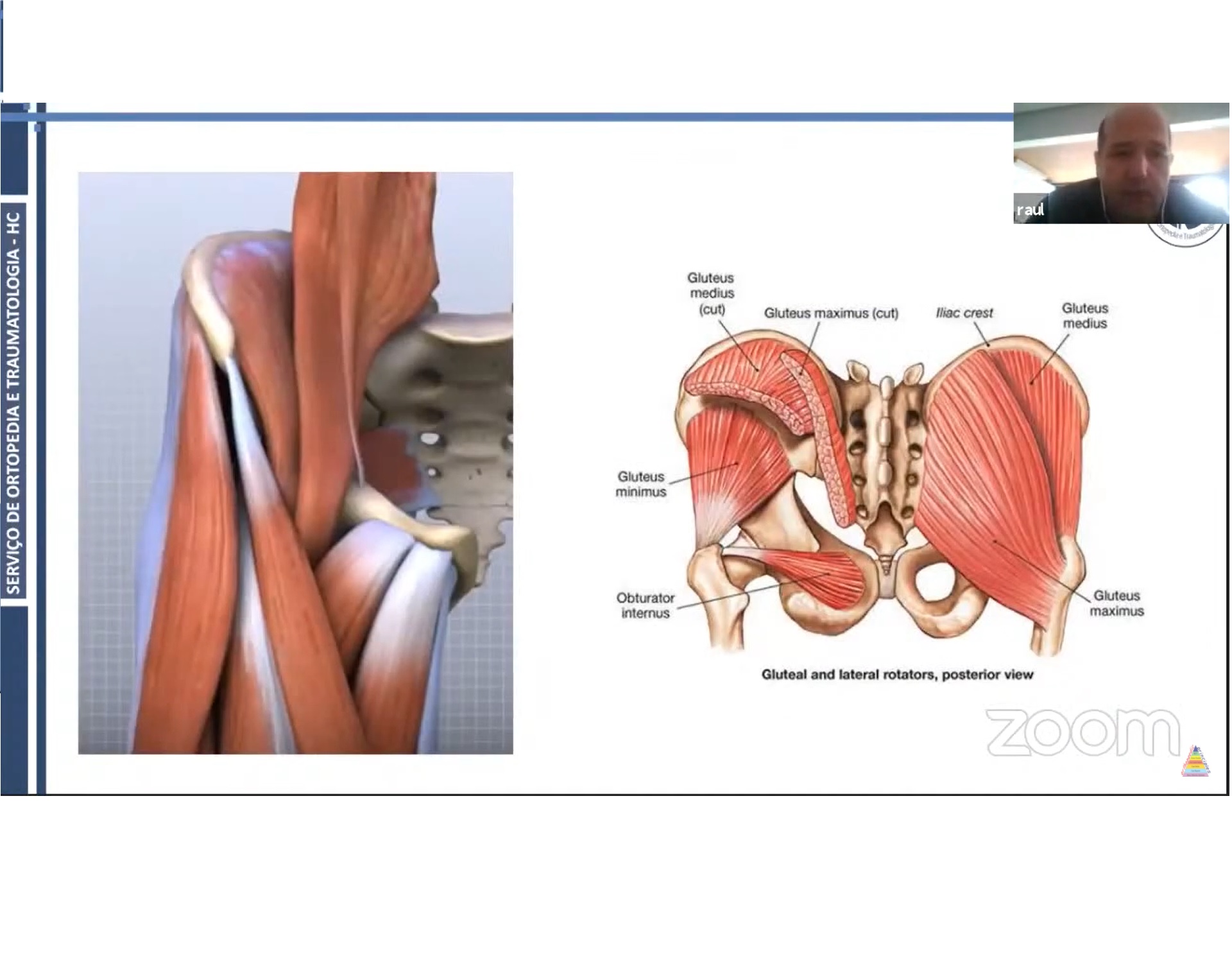
Anatomy of the Gluteus Medius
- The gluteus medius originates from the external surface of the ilium between the anterior and posterior gluteal lines.
- It inserts predominantly onto the lateral and superolateral aspects of the greater trochanter.
- The muscle contributes primarily to:
- Hip abduction.
- Pelvic stabilization during single-leg stance.
- Control of femoral and pelvic movement during gait.
- Different portions of the muscle have different functional roles, with contributions to hip rotation depending on the portion activated.
- The gluteus medius works together with:
- Gluteus minimus.
- Tensor fascia lata.
- Other muscles around the hip.
- The gluteus medius and minimus tendons have important relationships with the greater trochanter, trochanteric bursae and iliotibial band.
Biomechanical Importance
- The hip abductors play a major role in maintaining pelvic stability during walking and single-leg stance.
- During gait, the gluteus medius counteracts the tendency of the pelvis to drop toward the unsupported side.
- Weakness or failure of the abductors can therefore result in:
- Pelvic instability.
- Compensatory gait.
- Increased lateral hip loading.
- Trendelenburg gait.
- The iliotibial band also contributes to lateral hip stability and interacts mechanically with the greater trochanter and surrounding soft tissues.
- Persistent abductor dysfunction can significantly impair walking, stair climbing and athletic activity.
Epidemiology
- Greater trochanteric pain syndrome is common, particularly among middle-aged and older women.
- It is also associated with:
- Low back pain.
- Obesity.
- Hip osteoarthritis.
- Iliotibial band disorders.
- Altered gait mechanics.
- The prevalence of actual gluteus medius or minimus tears is considerably lower than the prevalence of the broader greater trochanteric pain syndrome.
Clinical Presentation
Typical symptoms include:
- Pain over the lateral aspect of the hip.
- Tenderness around the greater trochanter.
- Pain radiating down the lateral thigh.
- Difficulty lying on the affected side.
- Night pain or sleep disturbance.
- Pain while climbing stairs.
- Pain during prolonged walking or running.
- Difficulty with single-leg activities.
- Pain during resisted hip abduction.
- Possible associated low back pain.
Important point
- A Trendelenburg sign or gait abnormality is not necessary for the diagnosis.
- A patient with a partial gluteal tendon tear may have preserved gait and strength.
- Marked weakness or Trendelenburg gait increases suspicion for a more significant abductor lesion.
Physical Examination
Palpation
- Palpate the greater trochanter and surrounding structures.
- Reproduction of the patient’s characteristic pain with palpation supports the diagnosis of greater trochanteric pain syndrome.
- Tenderness should be correlated with other examination findings rather than interpreted in isolation.
Resisted Hip Abduction
- Pain or weakness during resisted hip abduction may indicate gluteal tendon pathology.
- Objective weakness is particularly important when evaluating suspected tendon tears.
Single-Leg Stance
- The patient is asked to stand on the affected limb.
- Pain reproduction supports a diagnosis of greater trochanteric pain syndrome.
- Pelvic dropping may indicate significant abductor dysfunction.
Trendelenburg Test
- The patient stands on one leg.
- A positive test occurs when the pelvis drops on the unsupported side.
- It suggests inadequate function of the hip abductors.
- A positive Trendelenburg test is particularly relevant in patients with substantial gluteal tendon tears.
Iliotibial Band Assessment
- The iliotibial band and tensor fascia lata should be assessed.
- Iliotibial band-related pathology may produce lateral hip or lateral thigh symptoms.
- External snapping hip should also be considered.
FABER Test
- Flexion, abduction and external rotation testing may help assess intra-articular hip pathology.
- It is not a specific test for gluteal tendon tears.
- A positive test should be interpreted in the context of the complete clinical examination.
Differential Diagnosis
Important alternative diagnoses include:
- Lumbar radiculopathy.
- Lumbar degenerative disease.
- Sacroiliac joint pathology.
- Hip osteoarthritis.
- Femoroacetabular impingement.
- Labral pathology.
- Iliotibial band syndrome.
- External snapping hip.
- Ischiofemoral impingement.
- Proximal hamstring pathology.
- Stress fracture.
- Hip abductor tendon tear.
- Trochanteric bursitis.
- Myofascial pain.
- Less commonly, neoplastic or infectious pathology.
Imaging
Plain Radiographs
- Plain radiographs are useful primarily for:
- Excluding intra-articular hip disease.
- Identifying osteoarthritis.
- Detecting fractures or other bony pathology.
- Identifying calcification around the greater trochanter.
- Detecting gross muscle wasting or other abnormalities.
- Plain radiographs cannot adequately characterize gluteal tendon tears.
Ultrasound
- Ultrasound is a useful investigation for peritrochanteric disorders.
- It can demonstrate:
- Gluteus medius tendinopathy.
- Gluteus minimus tendinopathy.
- Partial-thickness tears.
- Full-thickness tears.
- Tendon discontinuity.
- Calcification.
- Bursal abnormalities.
- Dynamic abnormalities such as snapping.
- Advantages include:
- Wide availability.
- Relatively low cost.
- Dynamic assessment.
- Comparison with the opposite side.
- Ability to guide injections.
- Important limitation:
- Ultrasound is operator-dependent.
- Tendon anisotropy can also create an appearance that mimics pathology if the probe is not positioned appropriately.
Magnetic Resonance Imaging
- Magnetic resonance imaging provides detailed assessment of:
- Gluteus medius tendon.
- Gluteus minimus tendon.
- Muscle quality.
- Fatty infiltration.
- Muscle atrophy.
- Peritrochanteric bursae.
- Adjacent soft tissues.
- Intra-articular hip pathology.
- MRI findings may include:
- Increased tendon signal.
- Tendon thickening.
- Partial-thickness tears.
- Full-thickness tears.
- Retraction.
- Peritendinous fluid.
- Muscle atrophy.
- Fatty degeneration.
- MRI is particularly useful when a significant tendon tear is suspected or when symptoms persist despite appropriate conservative treatment.
Important Imaging Principle
- Imaging abnormalities do not always correspond to symptoms.
- Abnormal tendon signal may be found in individuals without significant clinical symptoms.
- Therefore, MRI findings should always be correlated with:
- History.
- Physical examination.
- Functional impairment.
- Imaging should support the diagnosis rather than replace clinical reasoning.
Classification of Gluteal Tendon Lesions
Gluteal tendon pathology can be described according to:
Duration
- Acute.
- Chronic.
Extent
- Tendinopathy.
- Partial-thickness tear.
- High-grade partial-thickness tear.
- Full-thickness tear.
Muscle Quality
- Muscle atrophy.
- Fatty infiltration.
- Muscle quality is clinically important because chronic tendon dysfunction can result in irreversible muscle changes and may influence surgical outcomes.
Conservative Treatment
Most patients with greater trochanteric pain syndrome and gluteal tendinopathy can initially be treated nonoperatively.
Important components include:
- Education regarding the condition.
- Modification of aggravating activities.
- Avoidance of excessive compression of the gluteal tendons.
- Progressive hip abductor strengthening.
- Correction of movement patterns.
- Gait modification where necessary.
- Weight management where appropriate.
- Analgesic or anti-inflammatory medication when clinically appropriate.
- Gradual return to normal activity.
Exercise-Based Rehabilitation
- Education combined with targeted exercise has strong clinical support for gluteal tendinopathy.
- Rehabilitation should focus on:
- Progressive strengthening of the hip abductors.
- Improving pelvic control.
- Correcting excessive hip adduction during functional activities.
- Gradual progression of tendon loading.
- Restoration of functional capacity.
- A randomized clinical trial involving 204 patients demonstrated that education plus exercise produced better overall improvement than corticosteroid injection at long-term follow-up.
Corticosteroid Injection
- Corticosteroid injections may provide short-term symptom relief in selected patients.
- Their benefit may diminish with time.
- They should not replace appropriate rehabilitation.
- Repeated injections should be approached cautiously, particularly when significant tendon pathology is present.
Platelet-Rich Plasma
- Platelet-rich plasma has been investigated for gluteal tendinopathy.
- Evidence remains variable, with differences between studies in:
- Patient selection.
- Preparation methods.
- Injection protocols.
- Follow-up duration.
- It should therefore not be considered universally superior to structured rehabilitation.
Extracorporeal Shock-Wave Therapy
- Extracorporeal shock-wave therapy is another nonoperative option used in selected patients.
- Evidence suggests that it may provide benefit in chronic tendinopathy.
- It may be considered when symptoms persist despite appropriate education and exercise.
Indications for Surgery
Surgical treatment may be considered when there is:
- Persistent pain despite an adequate course of nonoperative treatment.
- Significant functional limitation.
- A high-grade partial-thickness or full-thickness tear.
- Objective abductor weakness.
- Significant gait abnormality.
- Failure of appropriate rehabilitation.
- A reparable tendon with acceptable muscle quality.
- The decision should not be based solely on the percentage of tendon involvement.
- Tear morphology, retraction, muscle atrophy, fatty infiltration, symptoms and functional impairment should all be considered.
Clinical studies have shown that reduced resisted-abduction strength and gait deviation are associated with a greater likelihood of proceeding to operative treatment in patients with gluteus medius tears.
Surgical Options
Surgery can be performed using:
- Open repair.
- Endoscopic repair.
Endoscopic Repair
Potential advantages include:
- Smaller incisions.
- Less soft-tissue disruption.
- Reduced postoperative morbidity.
- Ability to evaluate and treat associated peritrochanteric pathology.
- Potentially faster early recovery.
Potential technical challenges include:
- Limited working space.
- Portal placement.
- Identification of the tendon tear.
- Anchor placement.
- Management of large or retracted tears.
Open Repair
Open surgery may be preferred in selected cases, particularly when:
- The tear is extensive.
- The tendon is significantly retracted.
- Reconstruction is required.
- Tendon approximation is difficult.
- Muscle quality is poor.
- Tendon transfer is required.
Current evidence suggests that both open and endoscopic repair can produce meaningful clinical improvement in appropriately selected patients.
Principles of Endoscopic Repair
A typical endoscopic procedure involves:
- Positioning the patient appropriately.
- Establishing access to the peritrochanteric or subgluteal space.
- Bursectomy to improve visualization.
- Identification of the gluteal tendon.
- Assessment of the extent of tendon disruption.
- Identification of the normal tendon margins.
- Preparation of the greater trochanteric footprint.
- Placement of suture anchors.
- Tendon fixation to the prepared footprint.
- Confirmation of satisfactory tendon coverage and stability.
Assessment of Tendon Reparability
- Large chronic tears may be difficult to mobilize.
- Before committing to an endoscopic repair, the surgeon should assess whether the tendon can adequately cover its anatomical footprint.
- If adequate approximation is impossible, conversion to an open procedure or reconstruction may be required.
- Chronic tears with substantial retraction, muscle atrophy and fatty infiltration may require reconstructive strategies rather than simple repair.
Tendon Reconstruction
- When direct repair is not possible, reconstructive options may include:
- Tendon augmentation.
- Allograft or other graft reconstruction in selected cases.
- Gluteus maximus transfer.
- Other muscle-tendon transfer procedures.
- Gluteus maximus transfer is an option for selected irreparable abductor deficiencies.
- These procedures are technically demanding and should be reserved for carefully selected patients.
Double-Row Repair
- Double-row fixation may be used for selected larger tears.
- The objective is to improve tendon-to-bone contact across the greater trochanteric footprint.
- Multiple anchors may be used depending on:
- Tear size.
- Tendon quality.
- Bone quality.
- Footprint dimensions.
- Repair configuration.
- Anchor number and configuration should be individualized rather than applied as a fixed protocol.
Postoperative Care
Following substantial gluteal tendon repair:
- Protected weight bearing is commonly required.
- A hip brace may be used in selected patients.
- Crutches are used during the protected phase.
- Rehabilitation should be gradual.
- Excessive active abduction and tensile loading of the repair should be avoided during early healing.
- The exact duration of protection should depend on:
- Tear size.
- Repair technique.
- Tissue quality.
- Associated procedures.
- Surgeon-specific protocol.
Factors Associated With Poorer Outcomes
Potential adverse prognostic factors include:
- Advanced age.
- Chronic tears.
- Significant muscle atrophy.
- Fatty infiltration.
- Large or retracted tears.
- Poor tendon quality.
- Persistent abductor weakness.
- Delayed treatment in appropriate surgical candidates.
- However, these should be viewed as risk factors rather t

Leave a Reply