Courtesy: Shital Parikh, Taral Nagda, IORG, OrthoTV
Fracture Mimickers in Children (Pediatric Radiology Pitfalls)
1. Overview
- Pediatric X-rays often show normal variants mimicking fractures
- Common causes:
- Ossification centers
- Physeal anatomy
- Growth-related changes
Key Principle
- Always correlate with:
- Clinical findings
- Contralateral X-ray
2. Physiological Periosteal Reaction (Periostitis of Infancy)
Epidemiology
- Age: < 4 months
- Seen in ~30–35% of infants
Common Bones
- Tibia > Femur > Humerus
Features
- Diffuse periosteal reaction along shaft
Mimics
- Fracture
- Infection
Differentiation
- Periostitis:
- Diffuse
- Age < 4 months
- Fracture:
- Focal
- Any age
3. Cervical Spine Variants
A. Pseudosubluxation (C2–C3)
- Common in children
Diagnosis
- Spinolaminar line:
- Continuous – normal
- Disrupted – true subluxation
B. Atlanto-Dens Interval
- Children: </= 5 mm
- Adults: </= 3 mm
C. Prevertebral Soft Tissue
- Increased thickness:
- Normal in children
- Not always pathology
D. Ossification Centers
- Incomplete fusion – fracture-like lines
- Smooth, sclerotic margins – normal
4. Vertebral Wedging
- Mild wedging – physiological
Pathological If
-
20% anterior height loss
5. Elbow Ossification Centers (High-Yield)
Mnemonic: CRITOE
- Capitellum
- Radial head
- Internal epicondyle
- Trochlea
- Olecranon
- External epicondyle
Clinical Importance
- Avoid misdiagnosing ossification centers as fractures
Common Pitfalls
- Trochlea – irregular (looks fragmented)
- Radial head – multiple fragments (normal)
- Olecranon – multiple centers
Tip
- Always compare with opposite side
6. Osteochondritis Dissecans vs Normal Ossification
Age-Based Rule
- < 8 years – irregular ossification (normal)
-
12 years – likely OCD
- 8–12 years – gray zone
7. Epiphyseal Variants
A. Cleft Epiphysis
- Mimics fracture line
- Normal variant
B. Spurs (Distal Radius)
- Common misdiagnosis as fracture
C. Metacarpal Physis Location
- 2nd–4th – distal
- 1st metacarpal – proximal
D. Pseudoepiphysis
- Extra physis-like line
- Disappears with growth
8. Distal Tibia Variants
Features
- Epiphysis may be:
- Wedge-shaped
- Irregular
Mimics
- Tillaux fracture
Special Variant
- Poland’s hump / Lister’s tubercle irregularity
- Normal physeal undulations
Clinical Note
- Injury here – higher risk of growth arrest
9. Proximal Humerus Physis
Appearance Changes with Rotation
- External rotation – triangular
- Neutral – rectangular
- Internal rotation – altered contour
Pitfall
- May mimic fracture on single view
10. Fifth Metatarsal Apophysis
Normal
- Longitudinal orientation
Fracture
- Transverse line
11. Fat Pad Sign (Elbow)
Normal
- Small anterior fat pad
Abnormal
- Large anterior fat pad (“sail sign”)
- Posterior fat pad (always abnormal)
Indicates
- Occult fracture
- e.g., supracondylar fracture
12. Occult Fractures
May Not Be Visible Initially
Look For
- Fat pad sign
- Subtle cortical break
Complications
- Compartment syndrome
- Vascular injury
13. Accessory Ossicles
Common Locations
- Ankle
- Foot
- Wrist
Features
- Smooth
- Rounded
- Sclerotic margins
Fracture Features
- Sharp
- Irregular edges
Examples
- Os subfibulare / sub-tibiale
- Os peroneum
- Inferior patellar ossicles
14. Bipartite Variants
A. Bipartite Patella
- Superolateral location
- Normal
B. Bipartite Navicular
- Rare
- Usually treated conservatively
15. Supracondylar Spur
- Normal variant
- Not osteochondroma
- Does not point away from physis
16. Cortical Irregularities (Tumor Mimics)
Example
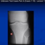
- Posteromedial distal femur irregularity
Cause
- Muscle traction:
- Adductor
- Gastrocnemius
Mimics
- Malignancy
17. Key Principles (Exam Pearls)
- Always correlate clinically
- Always compare with contralateral side
- Know CRITOE sequence
- Assess:
- Physis location
- Ossification timing
Important Rules
- Posterior fat pad = fracture until proven otherwise
- Smooth, corticated edges – normal variant
- Sharp, irregular edges – fracture
Final Message
- Most pediatric “fractures” on X-ray may actually be normal developmental variants
- Accurate interpretation requires:
- Knowledge of growth patterns
- Clinical correlation
- Careful radiological assessment
Leave a Reply