
Courtesy: Prof Ivan Wong, MD, FRCSC, Dalhousie University, Halifax, Canada
Advances in Hip Arthroscopy for Femoroacetabular Impingement
Overview
- Hip arthroscopy has evolved considerably with improvements in:
- Patient positioning.
- Ultrasound-guided access.
- Portal placement.
- Labral preservation and repair.
- Three-dimensional preoperative planning.
- Capsular management.
- Computer-assisted and augmented-reality technologies.
- These advances aim to improve access to the hip, optimize treatment of femoroacetabular impingement, reduce iatrogenic injury, and improve the accuracy of bony correction.
Lateral Decubitus Position for Hip Arthroscopy
- Hip arthroscopy can be performed in the lateral decubitus position, providing several practical advantages.
- Potential advantages include:
- Improved access to the medial aspect of the hip.
- Easier access to the posterior compartment.
- Greater working space around the operative field.
- Reduced interference between the surgeon and assistant.
- Elimination of the need for a C-arm in appropriately selected techniques.
- Smaller operating-room footprint.
- With modern postless traction systems, the patient can be stabilized without a perineal post.
- Postless positioning may reduce pressure-related complications associated with conventional perineal posts.
Ultrasound-Guided Hip Access
- Traditionally, fluoroscopy has been used to guide initial hip joint access.
- A typical fluoroscopic technique requires repeated imaging during needle advancement and portal establishment.
- Ultrasound provides real-time visualization of:
- The femoral head and neck.
- The acetabulum.
- The joint capsule.
- The advancing needle.
- This allows the surgeon to monitor the needle trajectory dynamically and avoid direct contact with the acetabular or femoral articular surfaces.
- Ultrasound-guided access can therefore facilitate hip arthroscopy without ionizing radiation.
Potential Benefits of Ultrasound Guidance
- Avoidance of radiation exposure.
- Real-time visualization of the needle.
- Potential reduction in repeated needle punctures.
- Potential reduction in inadvertent cartilage injury.
- Ability to perform hip access in operating rooms where fluoroscopy is unavailable.
- Three-dimensional spatial information can be obtained by manipulating the ultrasound probe.
Hip Arthroscopy Without Fluoroscopy
- Hip arthroscopy without routine fluoroscopy requires a detailed understanding of:
- Surface anatomy.
- Portal anatomy.
- Three-dimensional joint morphology.
- Instrument trajectory.
- Arthroscopic landmarks.
- Ultrasound can assist with accurate initial access.
- Once the joint has been entered, arthroscopic landmarks can guide subsequent procedures.
Acetabular Labrum: Functional Importance
- The acetabular labrum contributes significantly to normal hip biomechanics.
- Important functions include:
- Maintaining the suction seal around the femoral head.
- Contributing to hip stability.
- Helping maintain synovial fluid distribution.
- Participating in load transmission.
- Reducing local contact stresses.
- Helping maintain congruence between the femoral head and acetabulum.
- Preservation and repair of viable labral tissue are therefore important principles of contemporary hip preservation surgery.
Portal Selection in Hip Arthroscopy
- Several portals have been described for hip arthroscopy.
- Commonly used portals include:
- Anterolateral portal.
- Mid-anterior portal.
- Distal anterolateral accessory portal.
- Posterolateral portal.
- Medial or modified medial portals in selected cases.
- Portal selection should be individualized according to:
- Location and extent of the labral pathology.
- Acetabular morphology.
- Required instrument trajectory.
- Location of the planned anchor.
- Risk to the articular cartilage and surrounding neurovascular structures.
Anterolateral Portal
- Often serves as the initial viewing portal.
- Provides access to the central compartment and facilitates orientation within the joint.
- Establishing a consistent reference orientation is important for subsequent portal placement and acetabular work.
Mid-Anterior Portal
- Provides useful access to the anterosuperior acetabular rim.
- Can facilitate:
- Labral repair.
- Suture management.
- Anchor placement.
- Instrument manipulation around the anterior and anterosuperior rim.
Distal Anterolateral Accessory Portal
- Provides a different trajectory toward the acetabular rim.
- Its more distal and relatively parallel trajectory can be advantageous when the mid-anterior portal does not provide an appropriate angle for anchor insertion.
- Appropriate portal trajectory helps avoid penetration of the articular cartilage or subchondral bone.
Medial Portal
- Specialized medial access can be useful for pathology extending toward the 3 to 4 o’clock region of the acetabular rim.
- It may facilitate access to very medial labral tears and improve the trajectory for anchor placement.
- This is particularly relevant when the tear extends toward the transverse acetabular ligament.
- The portal should be established carefully because of the proximity of important neurovascular structures.
Posterolateral Portal
- Provides access to the posterior acetabular rim and posterior labrum.
- It may be useful when a labral tear extends substantially posteriorly.
- Appropriate portal selection can allow treatment of pathology around much of the circumference of the acetabular rim.
Principles of Labral Repair
- Successful labral repair requires:
- Adequate visualization.
- Appropriate portal placement.
- Accurate identification of the tear.
- Appropriate preparation of the acetabular rim.
- Secure anchor placement.
- Controlled suture passage.
- Restoration of the labral seal.
- Portal positioning should allow the surgeon to work around the labrum without damaging the articular cartilage.
- Medial or accessory portals may be required for tears that cannot be safely reached through conventional portals.
Femoroacetabular Impingement
- Femoroacetabular impingement is an important mechanical cause of hip pain and is associated with labral and cartilage pathology.
- The two major morphological patterns are:
- Cam morphology — abnormal prominence at the femoral head-neck junction.
- Pincer morphology — excessive or abnormal acetabular coverage.
- Mixed morphology is also common.
- Cam morphology can produce abnormal contact between the femoral head-neck junction and acetabular rim during hip movement.
- Repetitive mechanical conflict may contribute to:
- Labral injury.
- Chondral damage.
- Cartilage delamination.
- Progressive degenerative changes in susceptible patients.
Importance of Complete Cam Resection
- Accurate correction of cam morphology is one of the technically demanding components of hip arthroscopy.
- Both under-resection and over-resection should be avoided.
- Residual cam morphology is an important recognized cause of persistent symptoms and revision hip arthroscopy.
- The three-dimensional distribution of cam morphology may extend:
- Anterosuperiorly.
- Laterally.
- Inferiorly.
- Posterolaterally.
- A lesion that appears adequately treated on a standard anteroposterior radiograph may still have residual deformity in another region.
Imaging for Cam Morphology
Radiographs
- Common radiographic assessments include:
- Anteroposterior pelvis.
- Dunn views.
- Frog-leg lateral views.
- False-profile views when appropriate.
- Alpha angle and femoral head-neck offset are commonly used to assess cam morphology.
Magnetic Resonance Imaging
- Magnetic resonance imaging is particularly useful for evaluating:
- Labral pathology.
- Articular cartilage.
- Subchondral bone.
- Associated intra-articular pathology.
- Magnetic resonance arthrography can improve detection of certain labral abnormalities, although the choice between conventional magnetic resonance imaging and magnetic resonance arthrography depends on the clinical question and imaging quality.
Computed Tomography
- Computed tomography provides detailed assessment of osseous morphology.
- Three-dimensional computed tomography reconstructions can be particularly useful for:
- Defining the extent of cam morphology.
- Understanding its circumferential distribution.
- Planning the amount and location of bone resection.
- Assessing residual deformity after surgery.
Three-Dimensional Models in Cam Resection
- Three-dimensional printed patient-specific models provide a physical representation of the patient’s femoral morphology.
- They can assist the surgeon in identifying:
- The exact location of the cam prominence.
- The superior and inferior limits of the deformity.
- Posterior and posterolateral extensions.
- Areas at risk of residual impingement.
- The model can serve as a reference during arthroscopy.
Advantages of Three-Dimensional Models
- Improved appreciation of complex three-dimensional morphology.
- Better understanding of the location and extent of cam deformity.
- Ability to correlate preoperative imaging with arthroscopic anatomy.
- Assistance with determining the intended resection boundaries.
- Potential improvement in the consistency of cam correction.
- Opportunity to rehearse technically difficult procedures before surgery.
Residual Cam Morphology
- Residual cam morphology can be difficult to recognize intraoperatively.
- Two-dimensional radiographs may underestimate deformity located outside the standard projection.
- Three-dimensional reconstruction can demonstrate residual deformity more clearly.
- A physical three-dimensional model may further improve spatial understanding because it allows direct manipulation of the anatomy.
- Intraoperative comparison between the arthroscopic view and the three-dimensional model can help identify regions requiring additional correction.
Quantitative Assessment of Cam Resection
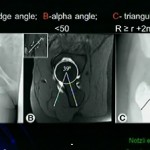
- The alpha angle is commonly used to assess femoral head-neck morphology.
- An alpha angle below approximately 55° has historically been used in many studies as a marker of satisfactory correction, although:
- Thresholds vary between studies.
- Measurement technique affects the value.
- No single radiographic measurement completely describes three-dimensional cam morphology.
- Evaluation should therefore incorporate the patient’s symptoms, clinical examination, multiple imaging views, and the overall three-dimensional morphology.
Surgical Technique: General Principles of Cam Resection
- The leg is positioned to provide optimal access to the femoral head-neck junction.
- Traction sutures may be used to improve visualization by retracting the capsule and surrounding soft tissues.
- The surgeon identifies the entire cam morphology rather than focusing on a single radiographic projection.
- Resection is performed progressively using an appropriate burr.
- The resection should follow the intended contour of the femoral head-neck junction.
- Multiple viewing positions and hip rotations can be used to inspect different regions.
- The goal is to restore an appropriate head-neck offset while preserving sufficient femoral neck strength.
- Excessive resection must be avoided because it can weaken the femoral neck and potentially increase the risk of fracture.
The “Double-Bump” Concept
- Complex cam morphology may have more than one prominent region.
- A second prominence can be missed if the surgeon relies on a single viewing angle.
- Recognition of this morphology is important because incomplete correction can leave clinically significant residual impingement.
- Three-dimensional imaging and patient-specific models can help identify these additional areas before and during surgery.
Three-Dimensional Planning and Surgical Outcomes
- Studies evaluating patient-specific three-dimensional models have reported improved consistency of radiographic cam correction compared with conventional imaging-based planning in selected settings.
- The potential explanation is that three-dimensional models allow the surgeon to understand the entire morphology rather than interpreting multiple two-dimensional images separately.
- Current evidence is encouraging, but three-dimensional printing should be regarded as an adjunct to established imaging and surgical techniques.
- Further clinical studies are required to establish whether improved radiographic correction consistently translates into superior long-term patient-reported outcomes and reduced revision rates.
Computer-Assisted and Augmented-Reality Planning
- Newer technologies are extending three-dimensional planning beyond printed models.
- Computer-generated three-dimensional maps can identify regions of potential femoroacetabular conflict.
- Augmented-reality systems may allow the surgeon to visualize a three-dimensional representation of the patient’s anatomy during surgery.
- Virtual anatomical models can potentially be manipulated to:
- Identify the position of the cam lesion.
- Correlate the virtual anatomy with the arthroscopic field.
- Define the desired resection boundary.
- Improve spatial orientation during surgery.
- These technologies remain developing tools and require appropriate validation before widespread adoption.
Diagnostic Intra-Articular Injection
- Diagnostic hip injection can be useful when the source of pain is uncertain.
- An intra-articular local anesthetic injection primarily helps determine whether symptoms are arising from the hip joint.
- It does not reliably distinguish every specific intra-articular pathology, such as a labral lesion versus acetabular dysplasia.
- When performing a diagnostic injection, assessment should include:
- Baseline pain.
- Pain during the patient’s usual provocative activities.
- Degree of pain relief after injection.
- Duration of relief while the local anesthetic is active.
- Specific movements or activities that become less painful.
- Ultrasound guidance can facilitate accurate intra-articular injection.
Acetabular Dysplasia and Diagnostic Reasoning
- Hip pain may arise from different structural abnormalities, including:
- Femoroacetabular impingement.
- Acetabular dysplasia.
- Labral pathology.
- Extra-articular disorders.
- Dysplasia represents a different biomechanical problem from cam-type impingement.
- In dysplasia, inadequate acetabular coverage can increase load transmission through the labrum and contribute to instability and overload.
- Treatment therefore requires careful assessment of acetabular morphology rather than treating an associated labral tear in isolation.
When Is Open Surgical Dislocation Considered?
- There is no universal cam-size threshold that mandates conversion from arthroscopic treatment to open surgical dislocation.
- The choice depends on:
- Location and extent of deformity.
- Associated structural abnormalities.
- Surgeon’s expertise.
- Previous surgery.
- Need for complex femoral head reconstruction.
- Availability of appropriate arthroscopic expertise and equipment.
- Open surgical dislocation provides extensive exposure of the femoral head and neck and can facilitate treatment of selected complex deformities.
- However, it is a technically demanding procedure.
- Preservation of the femoral head blood supply is critical because injury to the retinacular vessels can result in avascular necrosis.
- Detailed preoperative three-dimensional imaging can assist with planning either open or arthroscopic reconstruction.
Ischiofemoral Impingement
- Ischiofemoral impingement is an extra-articular cause of hip or buttock pain.
- It involves abnormal narrowing of the space between the ischium and the lesser trochanter, with potential irritation of the quadratus femoris and adjacent structures.
- Magnetic resonance imaging may demonstrate:
- Reduced ischiofemoral space.
- Quadratus femoris edema.
- Associated soft-tissue abnormalities.
- Diagnosis should integrate imaging findings with clinical examination because imaging abnormalities may not always be symptomatic.
Diagnostic Injection
- Ultrasound-guided diagnostic injection can help determine whether symptoms arise from the suspected extra-articular region.
- An intra-articular injection may also be useful when the differential diagnosis includes intra-articular pathology.
- The response to injection should be interpreted together with clinical findings and imaging rather than used as an isolated diagnostic test.
Subspine Impingement
- Subspine impingement is another form of extra-articular or extra-articular–intra-articular conflict around the anterior inferior iliac spine region.
- It may occur because of abnormal morphology or altered anatomy around the anterior acetabular region.
- Clinical assessment and appropriate imaging are important for identifying the source of symptoms.
- Treatment should be directed at the underlying structural abnormality only after establishing a convincing clinical diagnosis.
Key Takeaways
- Hip arthroscopy is increasingly supported by technologies that improve access, visualization, planning, and surgical precision.
- Lateral decubitus positioning can provide useful access to medial and posterior areas of the hip and can be combined with postless traction systems.
- Ultrasound-guided access allows real-time visualization of the needle and can facilitate hip arthroscopy without fluoroscopy.
- Appropriate portal selection is essential for safe and effective treatment of labral pathology.
- The acetabular labrum plays an important role in hip stability and the suction-seal mechanism and should be preserved whenever possible.
- Complete and appropriately contoured correction of cam morphology is critical because residual cam deformity is an important cause of persistent symptoms and revision surgery.
- Three-dimensional computed tomography and patient-specific three-dimensional models can improve understanding of complex cam morphology and assist with individualized surgical planning.
- Augmented-reality and computer-assisted three-dimensional technologies represent emerging developments in hip preservation surgery.
- Diagnostic intra-articular injection is useful for determining whether pain originates from within the hip joint, but it does not by itself establish the precise intra-articular diagnosis.
- Extra-articular conditions such as ischiofemoral and subspine impingement should be considered when symptoms and examination findings do not fit an isolated intra-articular disorder.
- Technology is most valuable when integrated with sound clinical reasoning, accurate diagnosis, detailed anatomical knowledge, and appropriate surgical expertise.
I need this complete paper
Excellent