
Courtesy: Prof Nabil Ebraheim, University of Toledo, Ohio, USA
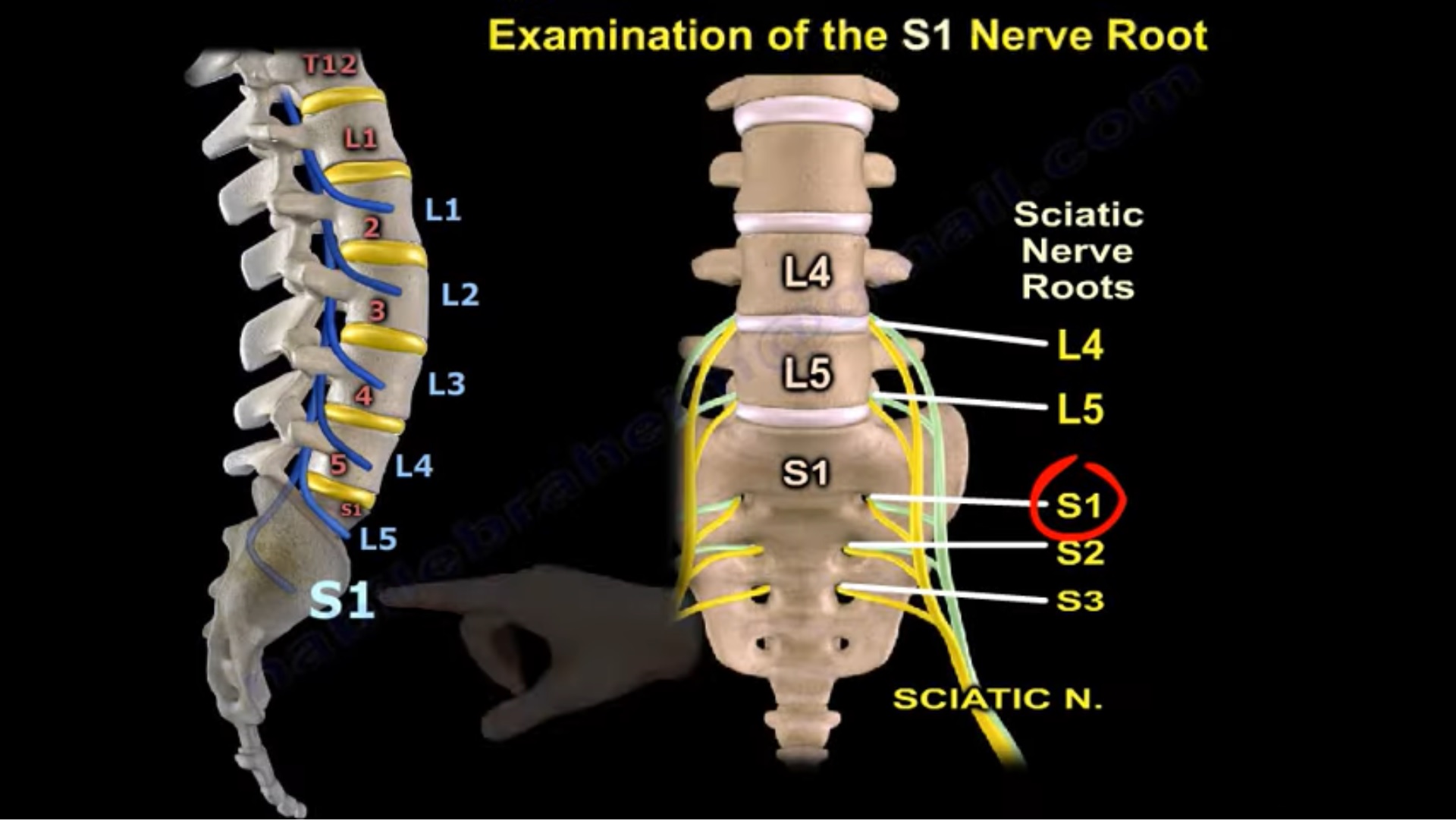
Examination of the S1 Nerve Root
Overview
S1 nerve-root examination into three parts:
- Sensory examination
2. Motor examination
3. Reflex examination
It also compares S1 with L5 and explains how different types of disc pathology at the L5–S1 level may affect different roots.
- Sensory Examination
For S1, examine sensation over the:
Lateral side of the foot
gives a useful comparison across the foot:
- L4 – medial side of foot
- L5 – dorsum/top of foot
- S1 l-ateral side of foot
Quick recall
Medial – L4
Top – L5
Lateral – S1
- Reflex Examination
The important reflex for S1 is:
Ankle reflex
So:
S1 – ankle jerk
- Motor Examination of S1
S1 motor examination is more complex because several muscles are supplied by the S1 root.
The muscles mentioned are:
- Gluteus maximus
- Lateral hamstrings
- Gastrocnemius
- Flexor hallucis longus
- Peroneus longus
- Peroneus brevis
- S1 Muscles From Proximal to Distal
from proximal to distal:
- Gluteus maximus
- Lateral hamstring
- Biceps femoris
- Gastrocnemius
- Flexor hallucis longus
- Peroneus longus and brevis
This gives a useful examination sequence from hip to foot.
- Important S1 Motor Functions
From the listed muscles, the main functions that can be assessed are:
| Muscle | Function relevant to examination |
| Gluteus maximus | Hip extension |
| Biceps femoris | Lateral hamstring function |
| Gastrocnemius | Plantar flexion |
| Flexor hallucis longus | Great-toe flexion |
| Peroneus longus/brevis | Foot eversion |
The does not provide detailed techniques for testing each individual muscle.
- EMG Findings in S1 Radiculopathy
A patient with:
L5–S1 disc herniation affecting the S1 root
may show EMG abnormalities in the S1-supplied muscles listed above.
The specifically mentions:
- Sharp waves
- Fibrillations
- L5–S1 Disc Herniation and Root Involvement
This is an important distinction in the .
Posterolateral L5–S1 Disc Herniation
Posterolateral L5–S1 disc herniation – S1 nerve-root irritation
This is the typical pattern emphasized by .
Foraminal L5–S1 Disc Herniation
In contrast:
Foraminal L5–S1 disc herniation – L5 radiculopathy
So the same disc level may affect a different root depending on the location of the herniation.
Key concept
Posterolateral L5–S1 – S1
Foraminal L5–S1 – L5
- L5–S1 Isthmic Spondylolisthesis
L5–S1 isthmic spondylolisthesis may cause L5 radiculopathy
This is contrasted with posterolateral L5–S1 disc herniation, which affects S1.
- Comparison With L5 Nerve Root
The lecture then contrasts S1 with L5.
L5 Motor Muscles Mentioned
- Extensor hallucis longus
- Extensor digitorum longus
- Medial hamstrings
- Gluteus medius
L5 Sensory Area
Dorsum/top of the foot
L5 Reflex
No specific reflex
Typical Disc Level
Posterolateral L4–L5 disc herniation ? L5 nerve-root involvement
S1 vs L5 — High-Yield Table
| Feature | L5 | S1 |
| Typical posterolateral disc level | L4–L5 | L5–S1 |
| Sensory area | Dorsum/top of foot | Lateral side of foot |
| Important distal muscle | EHL | Gastrocnemius |
| Additional muscles | EDL, medial hamstrings, gluteus medius | FHL, peronei, lateral hamstrings, gluteus maximus |
| Reflex | No specific reflex | Ankle reflex |
| Foraminal L5–S1 disc | Can affect L5 | — |
Disc Position and Root Affected
This is probably the most useful conceptual point in the :
At L5–S1
Posterolateral herniation
affects the traversing S1 root
Foraminal herniation
affects the L5 root
also associates:
L5–S1 isthmic spondylolisthesis – L5 radiculopathy
Practical S1 Examination Sequence
Sensory
Test:
Lateral side of foot
Motor
Examine S1-supplied muscles from proximal to distal:
Gluteus maximus – lateral hamstrings – gastrocnemius – FHL – peronei
Reflex
Check:
Ankle jerk
Then correlate with the suspected level and location of disc pathology.
Key Take-Home Points
- S1 examination consists of sensory + motor + reflex assessment.
- S1 sensory area – lateral foot.
- S1 reflex – ankle jerk.
- Important S1 muscles include:
- Gluteus maximus
- Biceps femoris
- Gastrocnemius
- Flexor hallucis longus
- Peroneus longus and brevis
- EMG in S1 radiculopathy may show fibrillation potentials/sharp waves in affected muscles.
- Posterolateral L5–S1 disc – S1 radiculopathy.
- Foraminal L5–S1 disc – L5 radiculopathy.
- L5–S1 isthmic spondylolisthesis – L5 radiculopathy according to .
- L5 has no specific reflex emphasized in this .
Exam Pearls
L4 – medial foot
L5 – dorsum of foot
S1 – lateral foot
S1 – plantar flexion + ankle jerk
L5–S1 posterolateral disc – S1
L5–S1 foraminal disc – L5
L4–L5 posterolateral disc – L5
Leave a Reply