
Courtesy: Prof Nabil Ebraheim, University of Toledo, Ohio, USA
Overview
-
Gout is a well-recognized cause of bursitis.
-
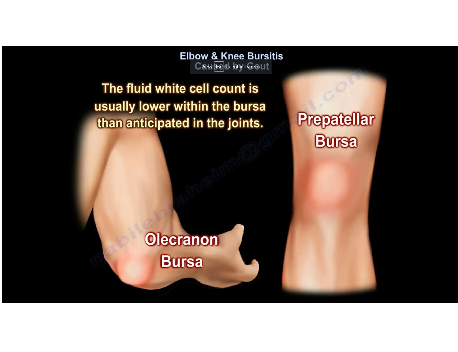
Gouty bursitis most commonly involves the olecranon bursa at the elbow and the prepatellar bursa at the knee.
Pathophysiology
-
In gouty bursitis, uric acid crystals are frequently present within the bursal fluid.
-
These crystals are needle-shaped and demonstrate negative birefringence under polarized light microscopy.
-
The presence of uric acid crystals within the bursa does not exclude concurrent infection.
Laboratory Findings
-
The white cell count in bursal fluid is usually lower than expected when compared with septic arthritis of a joint.
-
Despite lower cell counts, infection may still coexist and must be actively excluded.
Aspiration of the Bursa
-
Aspiration is essential for diagnostic evaluation.
-
Aspirated fluid should be sent for:
-
Culture and sensitivity
-
Cell count
-
Crystal analysis
-
Clinical Considerations
-
Gouty arthritis and bursitis may be bilateral.
-
These conditions can be difficult to treat, particularly in chronic or recurrent cases.
-
There is a recognized association between gouty bursitis and secondary infection, necessitating careful clinical and laboratory assessment.
Related Posts
 Bursitis of Knee
Courtesy: Prof Nabil Ebraheim, University of Toledo, Ohio, USA
 Rheumatology Pearls
Courtesy: John M. Davis, III, MD, Division of Rheumatology Mayo Clinic
- Bursitis around the Hip
Courtesy: Prof Nabile Ebraheim, University of Toledo, OH, USA
My physio today thinks the swollen bursae around my right knee is gout. Been in terrific pain for months. The right knee has a history of both ACL and meniscus tear going back 15 years and resurfaced three years ago jumping off the bed with leg twisted. At 81 and with a large bunion on the right foot I had enough issues…..now this. Anyway, being sent for a test of the bursa liquid and an MRI which I see is also recommended in this article. Thank you so much!