Courtesy: Saurabh Agarwal, Consultant, UK
Introduction
-
Perilunate injuries involve disruption of the ligamentous and bony relationships between the lunate, adjacent carpal bones, and the distal radius.
-
These injuries represent severe wrist trauma and can lead to persistent pain, instability, and functional impairment if not recognized early.
-
Perilunate and lunate dislocations together account for the majority of carpal dislocations.
Carpal Alignment and Gilula Lines
Assessment of carpal alignment on a posteroanterior wrist radiograph is performed using Gilula lines.
Gilula Lines
Three smooth arcs should normally be seen:
-
First arc
-
Formed by the proximal convex surfaces of the scaphoid, lunate, and triquetrum.
-
-
Second arc
-
Formed by the distal convex surfaces of the same bones.
-
-
Third arc
-
Formed by the proximal surfaces of the capitate and hamate.
-
Clinical Importance
-
Any break or irregularity in these arcs indicates possible carpal instability or fracture-dislocation.
Mechanism of Injury
Perilunate injuries usually occur following high-energy trauma.
Common causes include:
-
Fall on an outstretched hyperextended wrist
-
Motor vehicle accidents
-
Sports-related trauma
Pathomechanics
-
Force is transmitted through the extended wrist, producing progressive disruption of intercarpal ligaments and carpal alignment.
Stages of Perilunate Injury (Mayfield Classification)
The Mayfield classification describes the progressive sequence of ligament failure during wrist hyperextension injury.
Stage 1
-
Scapholunate ligament disruption
-
Results in scapholunate dissociation
Stage 2
-
Disruption between the capitate and lunate
-
Leads to lunocapitate dislocation
Stage 3
-
Disruption between the lunate and triquetrum
-
Produces perilunate dislocation, where the capitate displaces dorsally relative to the lunate.
Stage 4
-
Complete failure of stabilizing ligaments
-
The lunate dislocates volarly into the carpal tunnel
Types of Perilunate Injuries
Trans-Scaphoid Perilunate Dislocation
-
The most common pattern.
-
Characterized by:
-
Fracture of the scaphoid
-
Associated perilunate dislocation
-
Other Injury Patterns
-
Partial or complete lunate dislocation
-
Associated fractures involving:
-
Scaphoid
-
Triquetrum
-
Capitate
-
-
Independent displacement of fractured carpal fragments may occur.
Degrees of Lunate Displacement
Lunate dislocations can be graded according to the degree of rotation and displacement.
Grade 1
-
Normal lunate alignment.
Grade 2
-
Palmar rotation of approximately ninety degrees, but still attached to the radius.
Grade 3
-
Further rotation with partial loss of ligament attachment.
Grade 4
-
Complete enucleation of the lunate, with loss of attachment to the radius.
Clinical Presentation
Symptoms
Patients typically present with:
-
Severe wrist pain
-
Swelling
-
Visible deformity
-
Reduced wrist movement
Physical Findings
-
Radial deviation of the wrist
-
Tenderness around the carpus
-
Volar prominence of the lunate may be palpable
-
Markedly restricted wrist motion
Neurological Complication
-
Median nerve compression may occur due to volar displacement of the lunate into the carpal tunnel.
Diagnostic Imaging
Plain Radiographs
Radiographic evaluation should include posteroanterior and lateral wrist views.
Typical Findings
-
Disruption of Gilula arcs
-
Loss of normal carpal alignment
-
Associated fractures
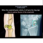
Classic Radiographic Signs
Lateral View
-
Spilled teacup sign
-
Represents volar rotation of the lunate.
-
Posteroanterior View
-
Triangular or piece-of-pie appearance of the lunate
-
Capitate not aligned with the lunate fossa of the radius
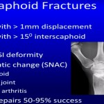
Associated Fractures
-
Scaphoid fracture
-
Triquetral fracture
Advanced Imaging
Computed Tomography
-
Provides detailed evaluation of carpal fractures and fragment displacement.
Magnetic Resonance Imaging
-
Useful for assessing ligament injuries and soft tissue damage.
Treatment
Management depends on severity, timing of diagnosis, and associated injuries.
Closed Reduction
Closed reduction may be attempted in acute cases.
Principle
-
Restoration of normal carpal alignment without surgery.
Technique
-
Apply longitudinal traction to the wrist.
-
Gradually flex the wrist while maintaining traction.
-
Manipulate the carpal bones to restore normal alignment.
Indicators of Successful Reduction
-
Restoration of Gilula arcs
-
Normal scapholunate gap
-
Proper intercarpal alignment
Immobilization
-
Wrist immobilized in a cast for approximately eight to twelve weeks.
Percutaneous Pinning
-
Kirschner wires may be used to stabilize:
-
Scapholunate joint
-
Lunotriquetral joint
-
Other unstable carpal relationships
-
This maintains alignment during ligament healing.
Surgical Management
Open Reduction and Internal Fixation
This is the preferred treatment in most cases, particularly when:
-
Closed reduction fails
-
Fractures are present
-
Instability persists
Surgical Goals
-
Restore carpal alignment
-
Repair injured ligaments
-
Fix associated fractures
-
Prevent long-term instability
Complications
Delayed diagnosis or inadequate treatment can result in:
-
Chronic carpal instability
-
Post-traumatic wrist arthritis
-
Median nerve injury
-
Persistent pain and reduced wrist motion
Outcomes
Functional outcome depends mainly on:
-
Early diagnosis
-
Accurate reduction
-
Stable fixation
-
Proper rehabilitation
Delayed treatment increases the risk of permanent wrist dysfunction and degenerative changes.
Key Points
-
Perilunate injuries represent serious wrist trauma requiring prompt recognition.
-
Radiographic evaluation using Gilula lines is essential for diagnosis.
-
The Mayfield classification describes progressive ligament injury stages.
-
Trans-scaphoid perilunate dislocation is the most frequent injury pattern.
-
Early reduction and stabilization significantly improve functional outcomes.

Leave a Reply