
Courtesy: Ramesh Sen, Fortis Hospital, Mohali
Avascular Necrosis of the Femoral Head: Diagnosis, Progression, and Treatment Strategies
Key Points
1. Overview of Avascular Necrosis
- Avascular necrosis is a condition caused by reduced blood supply to the femoral head.
- This leads to bone death and eventual collapse of the joint surface.
- The disease may progress gradually and symptoms may appear late.
2. Causes and Risk Factors
- Common causes include:
- Long-term steroid use
- Excessive alcohol intake
- Sickle cell disease
- In some regions, nutritional and metabolic factors may also contribute.
- Steroid dosage is strongly associated with increased risk.
3. Pathology
- In most cases, the condition is non-inflammatory.
- In specific conditions such as sickle cell disease, inflammatory changes may be present.
- The disease ultimately results in structural failure of the femoral head.
4. Clinical Presentation
- Pain is a common symptom but may appear late.
- Some patients may remain asymptomatic for several years despite disease progression.
- Physical examination may reveal limited hip movement and discomfort.
5. Imaging and Diagnosis
- X-rays:
- May be normal in early stages.
- Used for staging disease progression.
- Magnetic Resonance Imaging:
- Most sensitive tool for early detection.
- Shows characteristic signal changes in the femoral head.
- Helps determine the extent and location of involvement.
- Additional Imaging:
- Advanced imaging methods may assist in selected cases.
- Used for monitoring response to treatment.
6. Disease Progression
- The size and location of the lesion influence prognosis.
- Larger lesions are more likely to progress.
- Involvement of weight-bearing areas carries a poorer outcome.
- The condition may remain stable for years before becoming symptomatic.
7. Natural History
- Small lesions may remain stable over time.
- Larger lesions tend to progress or remain unchanged rather than improve.
- Joint collapse marks the transition to advanced disease.
8. Non-Surgical Management
- Restriction of weight-bearing alone does not significantly alter disease progression.
- Various medical therapies have been explored, including:
- Lipid-lowering agents
- Hyperbaric oxygen therapy
- Shockwave therapy
- Electromagnetic stimulation
- Anticoagulants in selected cases
- Bisphosphonates may help slow bone resorption, though evidence is variable.
9. Surgical Treatment Options
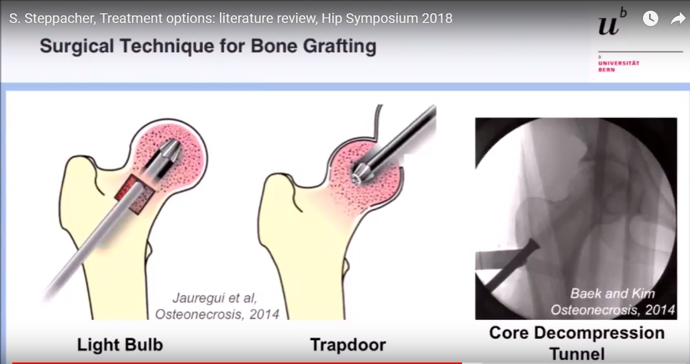
- Core Decompression:
- Standard early intervention.
- Reduces pressure within the bone and improves blood flow.
- More effective in early stages.
- Bone Grafting:
- Provides structural support and promotes healing.
- Can be combined with other techniques.
- Cell-Based Therapies:
- Bone marrow-derived cells may enhance healing.
- Increasing evidence supports their use in early disease.
- Vascularized Grafts:
- Restore blood supply to the affected area.
- Useful in selected patients.
- Osteotomy:
- Alters load distribution on the femoral head.
- Technically demanding and less commonly performed.
10. Advanced Disease Management
- Once collapse occurs, joint preservation becomes difficult.
- Total hip replacement is the most effective treatment in advanced stages.
- Previous surgeries may influence outcomes of joint replacement.
11. Treatment Approach by Stage
- Early stage:
- Core decompression, with or without adjunct therapies.
- Intermediate stage:
- Bone grafting or cell-based treatments.
- Advanced stage:
- Joint replacement surgery.
12. Key Takeaways
- Early diagnosis is critical to preserve the hip joint.
- Magnetic resonance imaging is essential for detecting early disease.
- Lesion size and location determine prognosis.
- Treatment should be stage-based and individualized.
- Joint replacement remains the definitive option in advanced cases.

Leave a Reply