
Courtesy: Prof Nabil Ebraheim, University of Toledo, Ohio, USA
Avascular Necrosis of the Femoral Head: Staging, Diagnosis, and Treatment Approach
Key Points
1. Overview
- Avascular necrosis is the death of bone tissue in the femoral head due to reduced blood supply.
- It is a progressive condition that can lead to collapse of the hip joint if untreated.
- The exact cause is not always clear, but several risk factors are known.
2. Risk Factors
- Risk factors can be broadly categorized into direct and indirect causes.
Direct Causes
- Trauma and radiation exposure.
- Blood-related disorders such as leukemia, lymphoma, and sickle cell disease.
- Metabolic and genetic conditions.
Indirect Causes
- Long-term steroid use.
- Excessive alcohol consumption.
- Chronic kidney disease.
- Organ transplantation.
- Autoimmune disorders.
- Coagulation disorders.
3. Bilateral Involvement
- Both hips may be affected in a large proportion of patients.
- The opposite hip should always be evaluated, even if there are no symptoms.
4. Importance of Early Diagnosis
- Early detection improves the success of joint-preserving procedures.
- Delayed diagnosis increases the risk of femoral head collapse.
5. Imaging and Diagnosis
- X-rays:
- May be normal in early stages.
- Later stages show structural changes and collapse.
- Magnetic Resonance Imaging:
- Most sensitive investigation for early disease.
- Shows characteristic signal changes in the femoral head.
Key Imaging Signs
- Crescent sign: indicates subchondral fracture.
- Double-line sign:
- Inner bright line represents vascular tissue.
- Outer dark line represents sclerotic bone.
6. Prognostic Factors
- Size of the lesion is the most important factor affecting progression.
- Smaller lesions with defined margins have better outcomes.
- Bone marrow edema indicates higher risk of progression.
7. Staging of Disease
- A commonly used staging system divides the disease into four stages:
Stage One
- Normal X-ray findings.
- Abnormal magnetic resonance imaging or bone scan.
Stage Two
- X-ray shows bone density changes.
- No collapse of the femoral head.
Stage Three
- Presence of crescent sign indicating subchondral fracture.
- Early collapse may be seen.
Stage Four
- Advanced collapse and deformity.
- Secondary degenerative changes in the joint.
8. Risk of Collapse
- Determined using combined necrotic angle measured on imaging.
- Larger angles are associated with higher risk of collapse.
9. Transient Osteoporosis
- A separate condition that can mimic osteonecrosis.
- Symptoms may be severe despite minimal changes on X-ray.
- Magnetic resonance imaging shows bone marrow edema.
- Does not require surgical treatment and usually resolves.
10. Treatment Strategies
- Early Stages (Before Collapse)
- Aim is to preserve the femoral head.
- Options include:
- Core decompression.
- Bone grafting or graft substitutes.
- Vascularized bone grafts.
- Use of biological therapies such as stem cells.
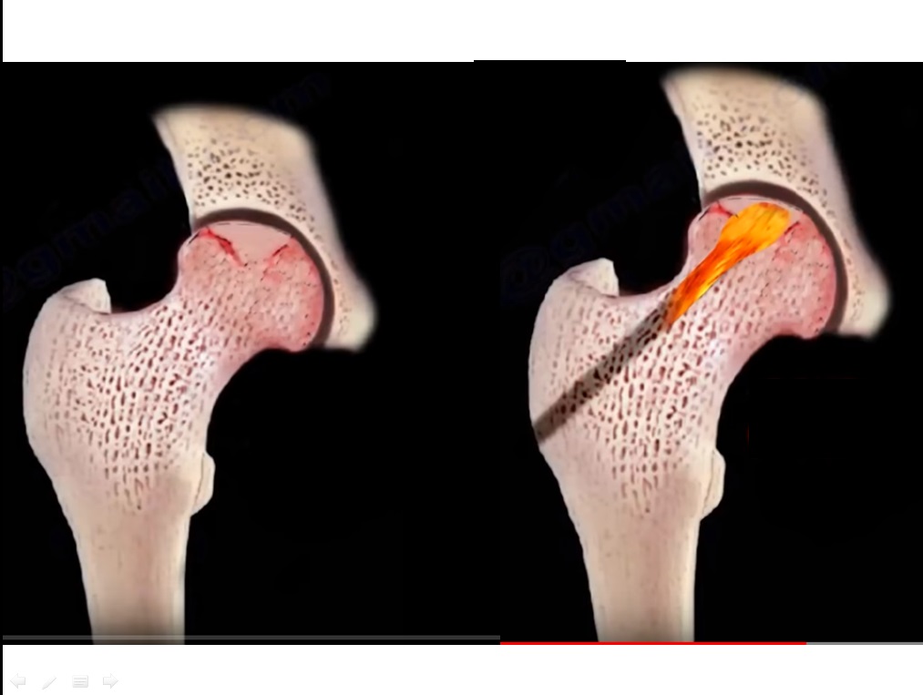
Core Decompression
- Involves drilling into the affected area to reduce pressure.
- Can be performed using a single large channel or multiple small channels.
- Promotes healing and reduces pain.
- Medical Management
- Bisphosphonates may reduce pain and risk of collapse.
- Their use is still considered under evaluation.
- Advanced Stages (After Collapse)
- Joint preservation is usually not possible.
- Total hip replacement is the treatment of choice.
- Provides reliable pain relief and improved function.
11. Surgical Options
- Bone Grafting Techniques
- Non-vascularized grafts.
- Vascularized fibular grafts, especially in younger patients.
- Total Hip Replacement
- Preferred in advanced disease.
- Cementless implants are commonly used.
12. Special Considerations
- In patients with sickle cell disease:
- Higher risk of disease progression.
- Surgical procedures may be technically more challenging.
- Younger patients may have slightly less favorable outcomes after joint replacement compared to those with osteoarthritis.
13. Key Takeaways
- Avascular necrosis is a progressive condition requiring early diagnosis.
- Magnetic resonance imaging is essential for early detection.
- Treatment depends on disease stage and severity.
- Joint-preserving procedures are effective in early stages.
- Total hip replacement is the definitive treatment in advanced cases.
Leave a Reply