• Skip to main content
  • Skip to secondary menu
  • Skip to primary sidebar
OrthopaedicPrinciples.com

OrthopaedicPrinciples.com

Integrating Principles and Evidence

Integrating Principles and Evidence

  • Home
  • Editorial Board
  • Our Books
    • Evidence Based Orthopaedic Principles
  • Courses
  • Exams
  • Reviews
  • Live Program
  • Contact

Arches of the Foot simplified

Courtesy: Sports Science Solutions, Arches of the Foot

Overview

The human foot is composed of multiple bones arranged to form structural arches.

These arches are essential for:

  • Strength and stability
  • Flexibility during movement
  • Shock absorption during walking and running

Key Functions of Foot Arches

  • Distribute body weight
  • Assist in propulsion during gait
  • Reduce impact forces on the lower limb

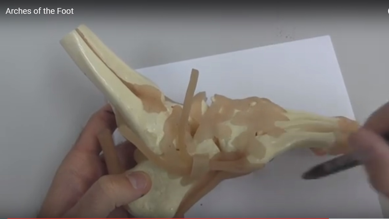
Bones Forming the Arches of the Foot

The arches are formed by a combination of tarsal and metatarsal bones:


Tarsal Bones

  • Calcaneus
  • Talus
  • Navicular
  • Cuboid
  • Three cuneiform bones:
    • Medial
    • Intermediate
    • Lateral

Metatarsal Bones

  • Five metatarsals

Types of Arches of the Foot

There are three main arches:

  1. Medial longitudinal arch
  2. Lateral longitudinal arch
  3. Transverse arch

Medial Longitudinal Arch


Structure

Extends from the heel to the forefoot on the medial side


Bones Involved

  • Calcaneus
  • Talus
  • Navicular
  • Three cuneiform bones
  • First three metatarsals

Keystone

  • Talus

 Represents the highest point of the arch


Characteristics

  • Higher and more elastic
  • Major role in:
    • Shock absorption
    • Weight distribution

Lateral Longitudinal Arch


Structure

Runs along the lateral side of the foot


Bones Involved

  • Calcaneus
  • Cuboid
  • Fourth metatarsal
  • Fifth metatarsal

Keystone

  • Cuboid

Characteristics

  • Lower and more rigid
  • Provides:
    • Stability
    • Support during standing and walking

Transverse Arch


Structure

Extends across the midfoot from medial to lateral side


Bones Involved

  • Cuboid
  • Three cuneiform bones
  • Bases of the metatarsals

Function

  • Maintains foot curvature across width
  • Helps in weight distribution across forefoot

Concept of the Keystone


Definition

The keystone is the central bone at the highest point of an arch, responsible for maintaining its structure and stability.


Examples

  • Talus — Medial longitudinal arch
  • Cuboid –Lateral longitudinal arch

Functional Importance of Foot Arches


1. Shock Absorption

  • Arches act as natural shock absorbers
  • Reduce transmission of impact forces

2. Spring Mechanism

  • Arches behave like a spring system:
    • Flatten under load — store energy
    • Recoil — release energy for propulsion

3. Weight Distribution

Body weight is distributed across:

  • Heel
  • Lateral border of the foot
  • Forefoot

4. Stability and Flexibility

  • Provide stability during standing
  • Allow flexibility during movement

Summary Points

  • The foot has three arches:
    • Medial longitudinal
    • Lateral longitudinal
    • Transverse
  • Talus — Keystone of medial arch
  • Cuboid — Keystone of lateral arch

Clinical Insight

  • Foot arches function as:
    • Shock absorbers
    • Energy-storing springs
  • The arrangement of multiple small bones allows:
    • Strength
    • Flexibility
    • Efficient weight transmission

Arches of the Foot simplified

Post Views: 2,547

Related Posts

  • Anatomy of the Sole of Foot

    Muscles of the plantar foot are divided into four layers: First layer: 3 muscles 1-Flexor…

  • Classification of Joints Simplified

    Courtesy: Sports Science Solutions, Lecture on Classification of Joints   Classification and Function of Joints…

  • Nerves of the Leg and Foot

    Courtesy: Prof Nabil Ebraheim, University of TOledo, Ohio, USA

Reader Interactions

Leave a Reply

Your email address will not be published. Required fields are marked *

Primary Sidebar

Follow Us

instagram slideshare

Categories

  • -Applied Anatomy
  • -Approaches
  • -Basic Sciences
  • -Cartilage & Meniscus
  • -Classifications
  • -Examination
  • -Foot and Ankle
  • -Foot and Ankle Trauma
  • -FRCS(Tr and Orth) tutorials
  • -Gait
  • -Hand and Wrist
  • -Hand and Wrist Trauma
  • -Hand Infections
  • -Hip and Knee
  • -Hip Preservation
  • -Infections
  • -Joint Reconstruction
  • -Knee Arthroplasty
  • -Knee Preservation
  • -Metabolic Disorders
  • -Oncology
  • -OrthoBiologics
  • -OrthoPlastic
  • -Paediatric Orthopaedics
  • -Paediatric Trauma
  • -Patellofemoral Joint
  • -Pelvis
  • -Peripheral Nerves
  • -Principles
  • -Principles of Surgery
  • -Radiology
  • -Rheumatology
  • -Shoulder and Elbow
  • -Shoulder and Elbow Arthroplasty
  • -Spine Deformity
  • -Spine Oncology
  • -Spine Trauma
  • -Spine, Pelvis & Neurology
  • -Sports Ankle and Foot
  • -Sports Elbow
  • -Sports Knee
  • -Sports Medicine
  • -Sports Medicine Hip
  • -Sports Shoulder
  • -Sports Wrist
  • -Statistics
  • -Technical Tip
  • -Technology in Orth
  • -Trauma
  • -Trauma (Upper Limb)
  • -Trauma Life Support
  • -Trauma Reconstruction
  • Book Shelf
  • Book Shelf Medical
  • Careers
  • Case Studies and Free Papers
  • DNB Ortho
  • Evidence Based Orthopaedic Principles
  • Evidence Based Orthopaedics
  • Exam Corner
  • Fellowships
  • Guest Editor
  • Guest Reviews
  • Image Quiz
  • Instructional Course Lectures
  • Journal Club
  • MCQs
  • Meetings and Courses
  • Multimedia
  • News and Blog
  • Plaster Techniques
  • Podcasts
  • Public Health
  • Rehabilitation
  • Research
  • Shorts and Reels
Copyright@orthopaedicprinciples.com. All right rerserved.