
Hip Arthroplasty Approaches & Surgical Hip Dislocation (Ganz Technique)
Part I: Comparative Hip Arthroplasty Approaches & Complications
Core Principle
-
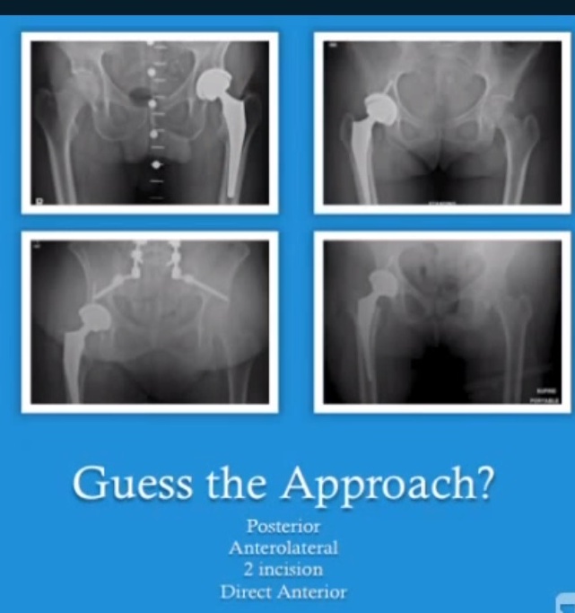
No Single Superior Approach: Postoperative radiographs from different approaches are largely indistinguishable; each approach involves trade-offs between visualization, stability, and approach-specific complications.
Overview of Arthroplasty Approaches
| Approach | Intermuscular / Interneural Plane | Key Advantages | Distinct Disadvantages / Complications |
| Trochanteric Osteotomy (Charnley) | None (bony osteotomy) | Broadest exposure; preserves medius/vastus lateralis continuity | High risk of non-union/hardware failure; rarely used for primary THA except in severe protrusio acetabuli |
| Posterior (Moore / Southern) | Gluteus maximus split; releases short external rotators & posterior capsule | Highly extensile (down to the knee); lowest intraoperative fracture rate; familiar anatomy | Higher dislocation risk historically; challenges with leg length/offset balance; 5× higher immediate post-op creatine kinase (CK) |
| Direct Lateral (Hardinge) | Gluteus maximus split; partial/total detachment of gluteus medius | Lower dislocation rate | Chronic abductor dysfunction (Trendelenburg lurch); superior gluteal nerve risk; denervation atrophy of TFL |
| Direct Anterior (DAA / Smith-Petersen) | True internervous plane: Sartorius (femoral n.) vs. TFL (superior gluteal n.) | Intermuscular/internervous; rapid early recovery; often no post-op hip precautions | High initial learning curve; intraoperative femur/calcar fractures; requires specialized traction table (or specialized technique); LFCN injury |
| Two-Incision Technique (MIs) | Dual portals: Anterior (acetabulum) & Posterior/Superior (femoral prep) | Low dislocation rate; potential for shorter hospitalization and lower acute inpatient cost | Non-extensile; specialized instrumentation required; steep learning curve; significant reports of heterotopic ossification (HO) |
Approach-Specific Controversies & Comparative Studies
-
Inflammatory Markers & Tissue Trauma (DAA vs. Posterior):
-
Creatine kinase (CK) levels were 5× higher in PACU and 2× higher cumulatively during hospitalization following posterior approaches.
-
Despite biomarker differences, blood loss and systemic recovery outcomes remained comparable between experienced cohorts.
-
-
The Two-Incision Debate:
-
Proponents: Reported shorter hospitalizations (discharge ~1 day earlier) and lower overall acute facility costs in select cohorts with BMI < 35.
-
Mayo Clinic / Pagano Series: Evaluated 3D gait analysis and isometric dynamometry, identifying no functional gait or strength benefit at 1 year over standard posterior approaches, alongside complications like severe heterotopic ossification requiring excision.
-
-
Direct Anterior Approach Complications:
-
In a series of 800 DAA cases over 5 years, elevated intraoperative fracture rates were observed early during the table-assisted learning curve.
-
A separate 200-case consecutive series on standard tables documented iatrogenic femur fractures and early dislocations driven by stem subsidence or unrecognized leg-length/offset discrepancies.
-
Inability to directly visualize or manage incidental abductor tears (present in ~20% of hip arthroplasty candidates).
-
Part II: Surgical Hip Dislocation (The Ganz / Trans-Trochanteric Flip)
Exposure & Advantages Over Kocher-Langenbeck
-
Supra-Acetabular Access: Extends visualization superiorly up to the anterior inferior iliac spine (AIIS).
-
360° Circumferential Joint Exposure: Direct visualization of the entire femoral head articular surface and the full acetabular perimeter (converting digital palpation into direct visual access).
-
Quadrilateral Surface Trade-Off: Access to the deep medial/quadrilateral plate is more constrained compared to a prone Kocher-Langenbeck approach, but this is offset by access to the superior dome.
Vascular Anatomy & Protection of Femoral Head Perfusion
-
Dominant Blood Supply: The ascending branch of the medial femoral circumflex artery (MFCA) provides terminal nutrient vessels penetrating the posterosuperior retinacular flap.
-
The Obturator Externus Cushion: The MFCA ascending branch passes directly anterior/superficial to the tendon of the obturator externus, which protects the vessel from stretch and crush injury during anterior hip dislocation.
-
Trochanteric Vascular Landmarks:
-
Trochanteric anastomosis running along the cranial border of the quadratus femoris (direct communication with the ascending branch).
-
Inferior gluteal anastomosis along the inferior border of the piriformis.
-
Surgical Technique: The Trochanteric Osteotomy Step-Cut
-
Osteotomy Design:
-
Flat Cut vs. Step-Cut: A stepped/chevron-type cut creates an inherent mechanical ledge that interdigitates upon reduction, providing baseline intrinsic stability prior to hardware fixation.
-
Preserving Tendinous Continuity: The posterior border of the gluteus medius tendon must retain a cuff of attached fibers to prevent notching of the femoral neck and protect ascending retinacular vessels (particularly in coxa vara or marked trochanteric overhang).
-
-
Gluteus Minimus Mobilization:
-
Gluteus minimus fibers attach further anteriorly and distally than traditionally described.
-
Muscular fibers lie near the superior gluteal neurovascular bundle; these fibers must be mobilized off the capsule to allow cephalad reflection without avulsion or ischemic injury.
-
-
Capsulotomy & Labral Management in Wall Fractures:
-
In posterior/superior wall fractures, the bone fragment must remain attached to its capsular pedicle to maintain blood supply.
-
If the fragment is tethered by an intact labrum that blocks visualization, a transverse radial cut through the labrum is performed to flip the fragment, followed by labral repair with suture anchors at closure.
-
Primary Indications
| Category | Specific Clinical Indications |
| Acute Acetabular Fractures | Posterosuperior and superior wall fractures requiring a direct 12:00 buttress plate out to the AIIS; Pipkin I/II femoral head fractures; Pipkin IV (combined head/wall) fractures |
| Complex Fractures | Select transverse, T-shaped, and transverse-posterior wall fracture patterns (historically requiring extended iliofemoral approaches) |
| Non-Arthroplasty Reconstructions | Post-traumatic malunions/non-unions; femoral head/neck osteochondroplasty for femoroacetabular impingement (FAI); osteochondral allograft plug (OATS) transfers |
| Pediatric / Adolescent Pathology | Chronic high-grade SCFE malunions (slip angle 65–70°); severe developmental dysplasia requiring combined reorientation and trochanteric advancement |
Technical Limitations & Pitfalls
-
Reduction Clamp Interference: The operative window is occupied by the dislocated femoral head, retinacular soft-tissue flap, and mobile abductor mass. Placing provisional reduction forceps can obstruct definitive plate and screw trajectories, requiring sequential reductions, temporary fixations, and re-dislocations.
-
Significant Intraoperative Blood Loss:
-
In the Siebenrock series (60 patients), blood loss averaged 1700 mL, primarily originating from the open, bleeding cancellous surfaces of the trochanteric osteotomy bed.
-
Mitigation: Seal the raw cancellous bone using bone wax, carboxymethylcellulose putty, or hemostatic agents.
-
Published Outcomes & Complications (Siebenrock 60-Patient Series)
-
Long-Term Joint Survival: Kaplan-Meier survival reached ~90% at 8 years for complex acetabular fracture reconstructions.
-
Avascular Necrosis (AVN): 0% incidence of AVN, demonstrating the vascular safety of preserving the MFCA ascending branch beneath the obturator externus.
-
Trochanteric Union: 100% bony union across the trochanteric osteotomy.
-
Nerve Injuries:
-
1 inferior gluteal nerve injury (resulting in tensor fasciae latae / abductor weakness).
-
1 femoral nerve palsy (secondary to a secondary anterior iliofemoral approach).
-
-
Heterotopic Ossification (HO): 4 cases of Brooker Class I; 1 case of Brooker Class II.
-
Author’s Personal Cautionary Complication: Severe, bridging Brooker Class IV heterotopic ossification in a 24-year-old with Reiter’s disease (ankylosing the joint and requiring extensive sciatic neurolysis).
Teaching Summary
-
Arthroplasty Takeaway: The “best” hip replacement approach is the one the surgeon has mastered; modern minimally invasive variants (DAA, two-incision) share similar long-term outcomes to conventional approaches but involve higher early technical complication curves.
-
Trauma Takeaway: The Ganz surgical hip dislocation provides circumferential visualization of the femoral head and superior acetabulum while preserving femoral head viability via the MFCA. However, it requires careful tissue mobilization and active hemostasis at the osteotomy site.
Courtesy: Dept of Orthopaedic Surgery, University of Washington, Seattle, Washington, USA
received mail today. thanks a lot. very much informative. -dr.k.d.shah.