Courtesy: Amr Abdelgawad, Maimonaides Medical Centre, Brooklyn, NY, USA
Medial Parapatellar Approach
- Most common approach for total knee arthroplasty.
- Incision runs medial to the patella.
- May injure medial genicular arteries (superior and inferior).
- Additional damage during lateral release or meniscectomy can compromise patellar blood supply.
- May result in avascular necrosis of the patella.
Subvastus and Midvastus Approaches
- Subvastus approach avoids quadriceps tendon incision.
- Provides less surgical exposure compared to medial parapatellar approach.
- Midvastus approach splits the vastus medialis muscle.
- Midvastus approach has higher risk of quadriceps denervation.
Extension of Exposure
- Quadriceps snip extends incision laterally from proximal end of medial parapatellar approach.
- Does not significantly change postoperative rehabilitation.
- V?Y turndown (patellar turndown) allows larger exposure.
- Patellar turndown requires delayed active extension postoperatively.
Posteromedial Knee Approach
- Used for bicondylar tibial plateau fractures and PCL tibial inlay graft.
- Interval between semimembranosus tendon and medial head of gastrocnemius.
- Medial gastrocnemius protects the neurovascular bundle.
- Popliteal artery approximately 2 cm from PCL tibial fixation screw.
Posterolateral Knee Approach
- Used for lateral meniscus inside?out repair.
- Superficial interval between iliotibial band and biceps femoris.
- Deep dissection achieved by retracting gastrocnemius posteriorly.
- Provides access to posterolateral capsule.
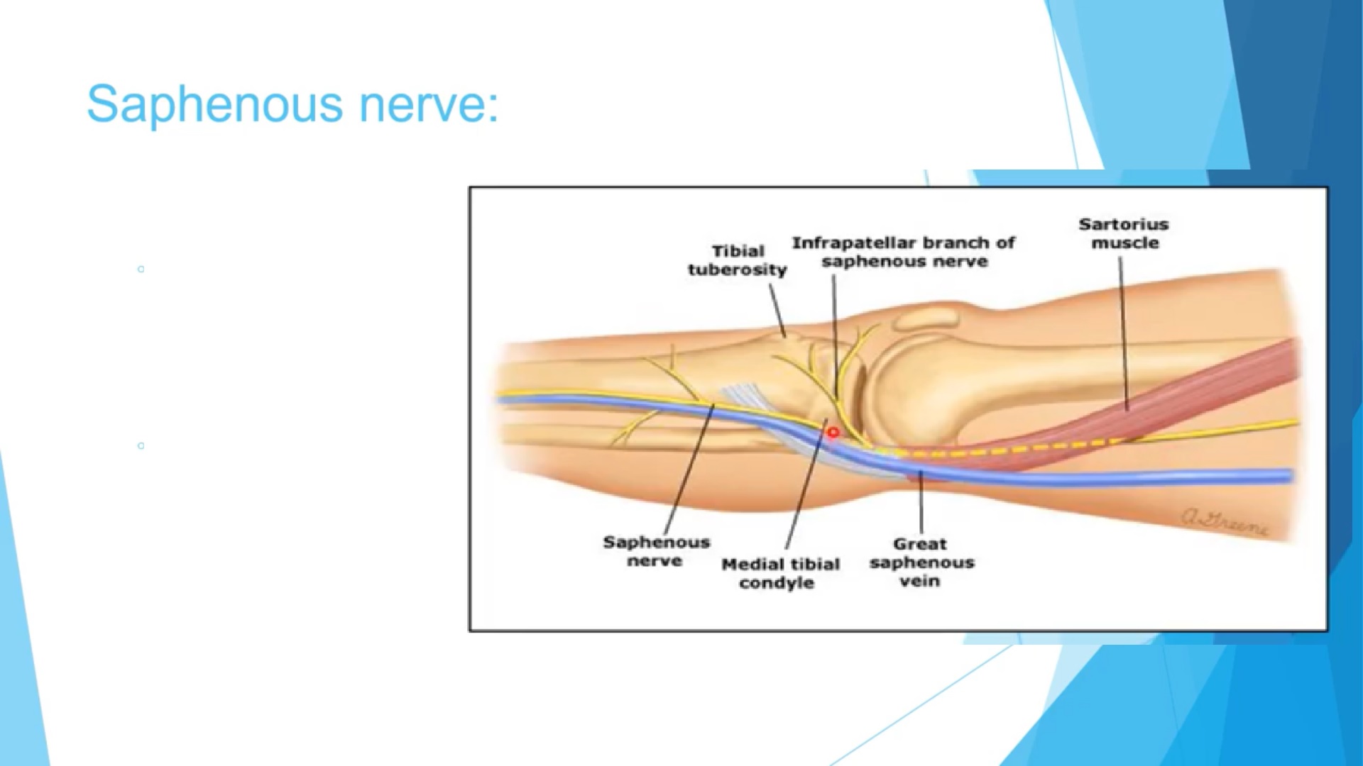
Saphenous Nerve at the Knee
- Infrapatellar branch crosses from medial to lateral.
- Midline incision (e.g., ACL BTB graft) may injure this branch.
- Injury causes numbness on lateral side of knee.
- Knee flexion and hip external rotation reduce nerve injury risk during hamstring graft harvest.
Lateral Knee Anatomy
- Popliteus tendon runs deep to the lateral collateral ligament.
- LCL lies proximal and posterior to popliteus tendon insertion.
- LCL inserts into anterior fibular head.
- Popliteofibular ligament inserts into posterior fibular head.
- Biceps femoris inserts broadly along fibular head.
Popliteal Neurovascular Bundle
- Arrangement superficial to deep: tibial nerve ? popliteal vein ? popliteal artery.
- Popliteal artery is the deepest and most anterior structure.
- Popliteal artery lies about 1 cm from posterior tibial plateau in flexion.
Screw?Home Mechanism
- Locks the knee in full extension.
- Occurs due to difference in femoral condyle geometry.
- External rotation of tibia occurs in final 20–30° of extension.
- Popliteus muscle unlocks the knee via internal rotation of tibia.
Blood Supply of the Knee
- Derived from popliteal artery.
- Includes superior genicular, inferior genicular, and middle genicular arteries.
- Middle genicular artery supplies ACL and PCL.
ACL Anatomy
- ACL is intra?articular but extrasynovial.
- Primary function: prevent anterior tibial translation.
- Secondary function: limit internal tibial rotation.
- Highest tension occurs in full extension.
- Blood supply from middle genicular artery.
ACL Bundles
- Two bundles: anteromedial (AM) and posterolateral (PL).
- AM bundle tight in flexion.
- PL bundle tight in extension.
- PL bundle provides rotational stability.
- Bundles separated by the lateral intercondylar ridge.
Meniscus Anatomy
- Menisci composed mainly of type I collagen.
- Lateral meniscus is more mobile than medial.
- Menisci move anteriorly with extension and posteriorly with flexion.
Baker’s Cyst
- Occurs between semimembranosus tendon and medial head of gastrocnemius.
- Often associated with intra?articular knee pathology.

Leave a Reply