Courtesy: Prof Nabil Ebraheim, University of Toledo, Ohio, USA
Classification
Unimalleolar Fracture
• Lateral malleolus fracture
• Medial malleolus fracture
• Posterior malleolus fracture
Bimalleolar Fracture
• Medial + lateral malleolus fractures
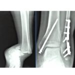
Trimalleolar Fracture
• Medial + lateral + posterior malleolus fractures
Important Point
• Obtain CT scan in trimalleolar fractures for:
• Posterior fragment size
• Fracture morphology
• Surgical planning
Key Concepts
Always assess for:
• Maisonneuve fracture
• Syndesmotic injury
• Deltoid ligament injury
• Stability of the ankle mortise
Ankle Stability
Most Important Stabilizer
• Deep deltoid ligament
Intact Deltoid Ligament
• Talus remains centered in mortise
• Stable ankle
Injured Deltoid Ligament
• Lateral talar shift
• Unstable ankle
Causes:
• Deltoid ligament rupture
• Medial malleolus fracture
Assessment on X-ray
Ask two questions:
1. Is the fracture displaced?
2. Is the fracture stable?
Stable Fracture
Typical example:
• Isolated undisplaced lateral malleolus fracture
• Talus remains congruent
Treatment:
• Walking boot or brace
• Weight bearing as tolerated
Stress Radiographs
Indication:
• Suspected deltoid ligament injury
Positive finding:
• Increased medial clear space
Indicates:
• Deep deltoid rupture
• Unstable ankle
Medial Clear Space
Normal
• ? 4 mm
• Or ? 2 mm greater than superior clear space
Abnormal
• > 4 mm
Suggests:
• Deltoid ligament injury
• Lateral talar shift
• Ankle instability
Supination External Rotation Injuries
SER Type II
• Stable
Treatment:
• Walking boot
• Progressive weight bearing
SER Type IV
• Deltoid ligament disruption
• Unstable
Treatment:
• Surgical fixation
Maisonneuve Fracture
Definition
• Proximal fibular fracture
• Syndesmotic disruption
• Unstable ankle injury
Clinical Pearls
• Often mistaken for ankle sprain
• Always palpate the entire fibula
• Obtain long leg radiographs
Treatment
• Syndesmotic reduction
• Syndesmotic screw fixation
Remember:
Maisonneuve Fracture = Syndesmotic Injury
High Fibular Fractures
Features
• Fibular fracture > 4.5 cm above ankle
Associated with:
• Syndesmotic disruption
• Increased requirement for syndesmotic fixation
Medial Malleolus Fractures
Tip Avulsion Fracture
• Usually treated nonoperatively
Typical Medial Malleolus Fracture
• Fix with two screws
Vertical Medial Malleolus Fracture
• Requires stronger fixation
Anterior Colliculus Fracture
• May require tension band wiring
Fibular Fixation
Standard technique:
- Interfragmentary lag screw
- Neutralization plate
Most commonly:
• One-third tubular plate
Order of Fixation
Standard sequence:
- Fibula
- Medial malleolus
- Posterior malleolus
- Assess syndesmosis
Exception:
• Comminuted fibula
Then:
• Fix medial malleolus first
Syndesmotic Injury
Assessment
Performed after fracture fixation using:
• Stress examination
• Cotton test
• Fluoroscopy
Radiographic Signs
Medial Clear Space
• > 4 mm abnormal
Tibiofibular Clear Space
• Normal < 5 mm
Syndesmotic Screw Fixation
Technique
• Screw inserted approximately 1.5 cm above ankle joint
Postoperative Care
• Non-weight bearing for 8 weeks
Screw Removal
Controversial:
• Some remove at 12 weeks
• Others retain screws
Evidence:
• Similar outcomes even with broken screws
Posterior Malleolus Fracture
Important Anatomy
• Posterior inferior tibiofibular ligament (PITFL) attaches to posterior fragment
Modern Concept
• Fragment morphology is more important than fragment size alone
Benefits of fixation:
• Restores syndesmotic stability
• Improves fibular reduction
• Improves ankle stability
Indications for Posterior Malleolus Fixation
• > 2 mm displacement
• Syndesmotic instability
• Large articular fragment
• Significant joint involvement
Failure to fix may cause:
• Syndesmotic instability
• Nonunion
• Post-traumatic arthritis
Posterior Malleolus Morphology
Type 1
Posterolateral Fragment
• Most common
Type 2
Shell Fragment
• Small fragment
• Often treated with syndesmotic fixation
Type 3
Large Posterior Fragment
• Partial articular injury
• Usually requires fixation
CT Scan
Essential for:
• Posterior fragment assessment
• Fracture morphology
• Surgical planning
X-rays frequently underestimate posterior injuries.
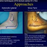
Posterior Approach
Posterolateral Approach
Most commonly used
Interval between:
• Flexor hallucis longus
• Peroneal tendons
Fixation:
• Buttress plate
Complications
• Syndesmotic malreduction
• Nonunion
• Post-traumatic arthritis
• Nerve injury
Most commonly injured nerve:
• Superficial peroneal nerve
Hardware Removal
• Routine implant removal is not mandatory
• Symptom improvement after removal is inconsistent
Exam Pearls
• Deep deltoid ligament is the key stabilizer of the ankle.
• Medial clear space > 4 mm indicates instability.
• Always examine the entire fibula in ankle injuries.
• Maisonneuve fracture is an unstable syndesmotic injury.
• CT scan is mandatory for trimalleolar fractures and posterior malleolar assessment.
• Posterior malleolar fixation improves syndesmotic stability.
• Fragment morphology is more important than fragment size alone.

Leave a Reply