• Skip to main content
  • Skip to secondary menu
  • Skip to primary sidebar
OrthopaedicPrinciples.com

OrthopaedicPrinciples.com

Integrating Principles and Evidence

Integrating Principles and Evidence

  • Home
  • Editorial Board
  • Our Books
    • Evidence Based Orthopaedic Principles
  • Courses
  • Exams
  • Reviews
  • Live Program
  • Contact

Anatomy Of The Upper Arm

Courtesy: Prof Nabil Ebraheim, University of Toledo, Ohio, USA

ANATOMY OF THE UPPER ARM
The muscles on the front of the upper arm consists of three muscles.
1. Biceps brachii muscle
2. Brachialis muscle
3. Coracobrachialis muscle

BICEPS BRACHII MUSCLE
• Biceps is a strong supinator of the forearm.
• Flexes the elbow joint.
• Long head of the biceps stabilizes the shoulder joint.

BRACHIALIS MUSCLE
• Primary flexor of the elbow joint.

CORACHOBRACHIALIS MUSCLE
• Flexion and adduction of the shoulder.
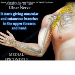
INNERVATION
• The musculocutaneous nerve arises from the lateral cord of the brachial plexus.
• It is the primary nerve supply to these 3 muscles.

Why it is musculocutaneous?
Musculo- It is the primary nerve supply of the muscles of the anterior compartment of the upper arm.
Cutaneous- It supplies sensation to the lateral half of the forearm.

TRICEPS MUSCLE:

• The triceps muscle is another muscle of the upper arm, but it is located on the back of the upper arm.

Function – Powerful extensor of the elbow joint.

Muscle has 3 heads :

1. Long head
2. Lateral head
3. Medial head

Innervation – Radial nerve from the posterior cord of the brachial plexus.

Post Views: 6,364

Related Posts

  • Acetabular fractures: Applied Anatomy

    Courtesy: Saqib Rehman MD Director of Orthopaedic Trauma Temple University Philadelphia Pennsylvania USA

  • Dorsal Wrist Anatomy

    Courtesy: Prof Nabil Ebraheim, University of Toledo, Ohio, USA

  • Ulnar Nerve Anatomy

    Courtesy: Prof Nabil Ebraheim, University of Toledo, Ohio, USA ORIGIN AND COURSE The ulnar nerve…

Reader Interactions

Leave a Reply

Your email address will not be published. Required fields are marked *

Primary Sidebar

Follow Us

instagram slideshare

Categories

  • -Applied Anatomy
  • -Approaches
  • -Basic Sciences
  • -Cartilage & Meniscus
  • -Classifications
  • -Examination
  • -Foot and Ankle
  • -Foot and Ankle Trauma
  • -FRCS(Tr and Orth) tutorials
  • -Gait
  • -Hand and Wrist
  • -Hand and Wrist Trauma
  • -Hand Infections
  • -Hip and Knee
  • -Hip Preservation
  • -Infections
  • -Joint Reconstruction
  • -Knee Arthroplasty
  • -Knee Preservation
  • -Metabolic Disorders
  • -Oncology
  • -OrthoBiologics
  • -OrthoPlastic
  • -Paediatric Orthopaedics
  • -Paediatric Trauma
  • -Patellofemoral Joint
  • -Pelvis
  • -Peripheral Nerves
  • -Principles
  • -Principles of Surgery
  • -Radiology
  • -Rheumatology
  • -Shoulder and Elbow
  • -Shoulder and Elbow Arthroplasty
  • -Spine Deformity
  • -Spine Oncology
  • -Spine Trauma
  • -Spine, Pelvis & Neurology
  • -Sports Ankle and Foot
  • -Sports Elbow
  • -Sports Knee
  • -Sports Medicine
  • -Sports Medicine Hip
  • -Sports Shoulder
  • -Sports Wrist
  • -Statistics
  • -Technical Tip
  • -Technology in Orth
  • -Trauma
  • -Trauma (Upper Limb)
  • -Trauma Life Support
  • -Trauma Reconstruction
  • Book Shelf
  • Book Shelf Medical
  • Careers
  • Case Studies and Free Papers
  • DNB Ortho
  • Evidence Based Orthopaedic Principles
  • Evidence Based Orthopaedics
  • Exam Corner
  • Fellowships
  • Guest Editor
  • Guest Reviews
  • Image Quiz
  • Instructional Course Lectures
  • Journal Club
  • MCQs
  • Meetings and Courses
  • Multimedia
  • News and Blog
  • Plaster Techniques
  • Podcasts
  • Public Health
  • Rehabilitation
  • Research
  • Shorts and Reels
Copyright@orthopaedicprinciples.com. All right rerserved.