Courtesy: Prof Nabil Ebraheim, University of TOledo, Ohio, USA
Overview
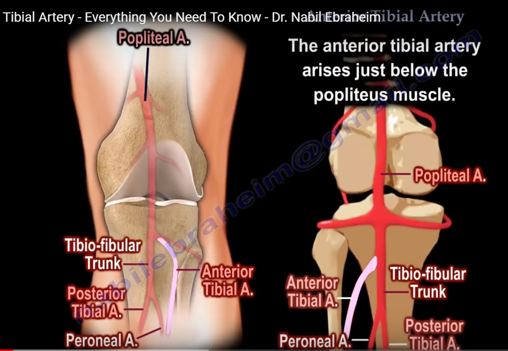
- The anterior tibial artery is one of the terminal branches of the popliteal artery.
- It supplies the anterior compartment of the leg and continues distally as the dorsalis pedis artery.
- Understanding its course and anatomical relationships is important in trauma, orthopedic surgery, and vascular assessment.
Origin of the Anterior Tibial Artery
- The popliteal artery divides into two major branches:
- Anterior tibial artery
- Posterior tibial artery
- In some individuals, the division occurs as:
- Anterior tibial artery
- Tibioperoneal trunk
- The tibioperoneal trunk then divides into:
- Posterior tibial artery
- Peroneal artery
Course of the Anterior Tibial Artery
- The artery arises just below the popliteus muscle.
- It passes through the interosseous membrane to enter the anterior compartment of the leg.
- It then descends along the anterior aspect of the leg toward the ankle.
Branches
The anterior tibial artery gives off important branches including:
- Anterior tibial recurrent artery
- Posterior tibial recurrent artery
Clinical Importance
- The anterior tibial recurrent artery may be injured in tibial tubercle fractures in children.
- Such injury can contribute to compartment syndrome of the leg.
Relations in the Leg
Proximal Leg
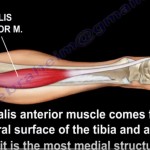
- The anterior tibial artery lies between:
- Tibialis anterior muscle (medially)
- Extensor digitorum longus muscle (laterally)
Middle Leg
- The extensor hallucis longus muscle appears between these muscles.
- At this level the artery lies between:
- Tibialis anterior
- Extensor hallucis longus
Distal Leg
- The extensor hallucis longus tendon crosses medially toward the great toe.
- The artery then lies between:
- Extensor hallucis longus (medially)
- Extensor digitorum longus (laterally)
Arrangement of Structures at the Anterior Ankle
At the level of the ankle joint, the structures are arranged from medial to lateral.
A common memory aid is:
Tom Has Very Nice Dog
Meaning:
- T – Tibialis anterior tendon
- H – Extensor hallucis longus tendon
- V – Vessels (anterior tibial artery)
- N – Deep peroneal nerve
- D – Extensor digitorum longus tendon
Important note:
- This arrangement applies mainly at the distal leg and anterior ankle, not in the proximal or middle portions of the leg.
Dorsalis Pedis Artery
- After passing beneath the extensor retinaculum, the anterior tibial artery becomes the dorsalis pedis artery.
- The dorsalis pedis artery supplies the dorsum of the foot and is an important site for palpation of peripheral pulses.
Deep Peroneal Nerve
- The deep peroneal nerve accompanies the anterior tibial artery.
- It enters the anterior compartment by piercing the intermuscular septum.
- It passes through the extensor digitorum longus muscle.
Relationship with the Artery
The position of the nerve changes along the course of the artery:
- Proximally: nerve lies lateral to the artery
- Middle part: nerve crosses anterior to the artery
- Distally: nerve again lies lateral to the artery
Radiological and Surgical Importance
- Imaging such as computed tomography angiography around the knee can demonstrate the branching pattern of the popliteal artery.
- At the level of the distal femur and proximal tibia, the following arteries may be visualized:
- Anterior tibial artery
- Posterior tibial artery
- Peroneal artery
Surgical Considerations
- During orthopedic procedures around the proximal tibia, careful placement of retractors is necessary.
- Improper retractor placement may damage the branches of the popliteal artery.
- This is particularly important because the bifurcation of the popliteal artery occurs in this region.
Summary Points
- The anterior tibial artery arises from the popliteal artery below the popliteus muscle.
- It passes through the interosseous membrane to enter the anterior compartment of the leg.
- It runs with the deep peroneal nerve and continues as the dorsalis pedis artery at the ankle.
- The anterior ankle structures follow the Tom Has Very Nice Dog arrangement from medial to lateral.
- Knowledge of this anatomy is essential for vascular assessment, trauma management, and orthopedic surgery.

Leave a Reply