Courtesy: Prof Nabil Ebraheim, University of Toledo, Ohio, USA
Overview
The patella is the largest sesamoid bone in the human body.
It is embedded within the quadriceps tendon and forms a crucial part of the knee extensor mechanism.
Key Functions
- Facilitates knee extension
- Enhances force transmission
- Protects the anterior aspect of the knee joint
Attachments of the Patella
Quadriceps Tendon
- Proximal attachment of the quadriceps muscle group
- A portion of the tendon extends over the anterior surface of the patella
Patellar Tendon
- Continuation of the quadriceps tendon
- Attaches the patella to the tibial tuberosity
Structure of the Patella
Apex
- Distal pointed portion
- Non-articular
- Attachment site for the patellar tendon
Articular Surface
- Located on the posterior surface
- Covered with thick articular cartilage (~5 mm centrally)
- Among the thickest cartilage in the body
Facets of the Patella
Main Articular Facets
- Medial facet
- Lateral facet
Lateral Facet
- Larger and wider
- Occupies the majority of the articular surface
Medial Facet
- Smaller (approximately half the size of the lateral facet)
Subdivisions
- Medial facet proper
- Odd facet
Odd Facet
- Located at the distal medial patella
- Articulates with the femur during deep knee flexion
Separation of Facets
- Divided by a vertical ridge
Bursae Around the Patella
Key bursae that reduce friction:
- Suprapatellar bursa
- Prepatellar bursa
- Infrapatellar bursa
Function of the Patella
Role in Knee Extension
- Acts as a pulley for the quadriceps mechanism
- Works with:
- Quadriceps muscle
- Patellar tendon
Mechanical Advantage
- Increases the moment arm of the quadriceps
- Moves the tendon away from the joint axis
- Improves efficiency of knee extension
Forces Acting on the Patella
Stabilizing Structures
Medial Stabilizers
- Medial retinaculum
- Vastus medialis
Lateral Stabilizers
- Lateral retinaculum
- Vastus lateralis
- Iliotibial band
Patellofemoral Joint Forces
- Patella engages in the trochlea at 40–45° of flexion
- Forces may reach 3–5× body weight during activity
Clinical Importance
Extensor Mechanism Injuries
Components
- Quadriceps muscle
- Quadriceps tendon
- Patella
- Patellar tendon
Key Consequence
- Disruption leads to loss of active knee extension
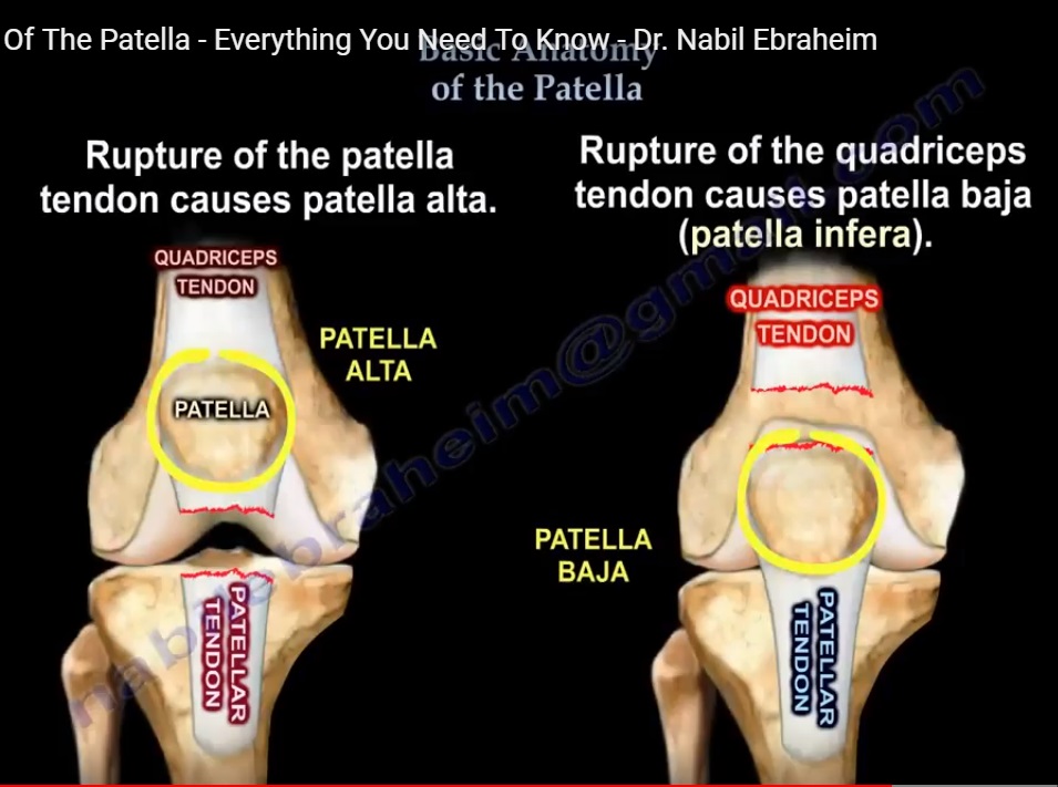
Patellar Tendon Rupture
- Quadriceps pulls patella upward
- Results in patella alta
Quadriceps Tendon Rupture
- Patellar tendon pulls patella downward
- Results in patella baja (infra)
Diagnosis
Clinical
- Often sufficient for complete tears
Imaging
- X-ray
- MRI (for confirmation and soft tissue evaluation)
Complications After Patellar Fracture Fixation
- Most common: Painful hardware
Medial Patellofemoral Ligament (MPFL)
Anatomy
- Inserts into the upper half of the medial patella
Function
- Primary restraint against lateral patellar displacement
Patellar Instability
Causes
- Injury to:
- Medial retinaculum
- Medial patellofemoral ligament
Result
- Lateral subluxation or dislocation of the patella
Imaging Findings
Sunrise (Axial) View
- Shows lateral displacement of patella
MRI Findings
- Bone bruising pattern:
- Medial patella
- Lateral femoral condyle
Summary Points
- Patella is the largest sesamoid bone and vital to the extensor mechanism
- Contains medial and lateral facets, separated by a vertical ridge
- Lateral facet is larger; medial facet includes the odd facet
- Enhances quadriceps efficiency
- Tendon injuries alter patellar position:
- Patella alta – patellar tendon rupture
- Patella baja – quadriceps tendon rupture
- MPFL is the key stabilizer preventing lateral dislocation
Leave a Reply