Courtesy: Rishi Dhir, London Hand and Wrist Course
FRCS Trauma Viva: Structure and High-Yield Cases
Structure of the Trauma Examination
Two Core Components
1. Decision-Making Component
Focuses on:
- Diagnosis
- Initial management
- Risk assessment
- Indications for surgery
2. Operative Component
Covers:
- Surgical techniques
- Approaches
- Complication management
Key Objective
Assess whether the candidate is safe to practice as a day-one consultant
Case 1: Fight Bite Injury (Clenched Fist Injury)
Clinical Scenario
- 24-year-old male
- Injury over dorsal MCP joint after a fight
Diagnosis
Fight bite injury
Key Features
- Tooth mark over MCP joint
- Occurs when fist strikes teeth
Structures Involved
- Zone V extensor mechanism
- Extensor tendon
- Sagittal band
- MCP joint capsule
Common Organisms
- Staphylococcus aureus
- Eikenella corrodens (exam favourite)
Assessment
History
- Time since injury
- Mechanism
- Tetanus status
- Comorbidities
Examination
- Neurovascular status
- Extensor tendon function
- Joint involvement
Investigations
- X-ray – fracture / foreign body
Tendon Assessment
Tenodesis Effect
- Wrist extension – finger flexion
- Wrist flexion – finger extension
– Loss suggests tendon rupture
Initial Management
- Tetanus prophylaxis
- IV antibiotics
- Irrigation
- Immobilization
Operative Management
Indication
Always requires surgical washout
Key Principles
- Position hand in clenched fist
- Open joint capsule (even if intact)
- Do NOT close wound primarily
Postoperative Plan
- Leave wound open
- Second look at 24–48 hours
Case 2: Pyogenic Flexor Tenosynovitis
Clinical Scenario
- Carpenter with splinter injury
- Severe finger pain
- Systemic symptoms
Diagnosis
Surgical emergency
Kanavel Signs
- Fusiform swelling
- Tender flexor sheath
- Pain on passive extension
- Finger held in flexion
Management
Initial
- IV antibiotics
- Splinting
- X-ray (foreign body)
Definitive
Surgery within 6 hours
Surgical Technique
Two-Incision Technique
- A1 pulley
- A5 pulley
Procedure
- Open sheath
- Insert cannula
- Irrigate
Advanced Infection
- Bruner zig-zag incision
Full exposure
Special Note
- Thumb & little finger – communicate with Parona’s space
Risk of horseshoe abscess
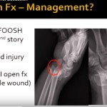
Case 3: Distal Radius Fracture Complication
Presentation
- Loss of thumb extension after fracture
Diagnosis
Extensor pollicis longus (EPL) rupture
Treatment
Extensor indicis proprius (EIP) tendon transfer
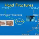
Case 4: PIP Joint Dislocation
Types
Simple
- No fracture
- Reducible
Complex
- Associated fracture
- Difficult reduction
Dorsal Dislocation (Most Common)
- Associated with volar plate injury
Treatment
- Closed reduction
- Early mobilization
Volar Dislocation
- Associated with central slip rupture
Treatment
- Extension splint
- ~6 weeks
MCP Joint Dislocation
Classification
- Simple – reducible
- Complex – irreducible
Cause of Irreducibility
Volar plate interposition
Key Principle
Do NOT apply traction
Reduction Technique
- Hyperextension
- Direct pressure
Case 5: Bennett vs Rolando Fractures
Bennett Fracture
Definition
- Intra-articular fracture at base of first metacarpal
Features
- Single fracture line
- Volar fragment attached to ligament
Treatment
- Closed reduction
- Percutaneous K-wires
Rolando Fracture
Definition
- Comminuted intra-articular fracture
Pattern
- Y-shaped
Treatment
- ORIF
- Mini-plates or K-wires
Surgical Approaches
Dorsal Approach
- Standard approach
Wagner Approach
- Junction of dorsal and glabrous skin
- Good access to volar fragments
Exam Strategy for FRCS Trauma Viva
Demonstrate
- Safe decision-making
- Recognition of emergencies
- Structured approach
Frequently Tested Topics
- Fight bite injury
- Flexor tenosynovitis
- EPL rupture
- Finger dislocations
- Bennett vs Rolando fractures
Key Take-Home Messages
- Always identify surgical emergencies early
- Avoid common pitfalls (e.g., traction in MCP dislocation)
- Use structured clinical reasoning
- Combine:
- Diagnosis
- Initial management
- Definitive treatment

Leave a Reply