Courtesy: Prof Nabil Ebraheim, University of Toledo, Ohio, USA
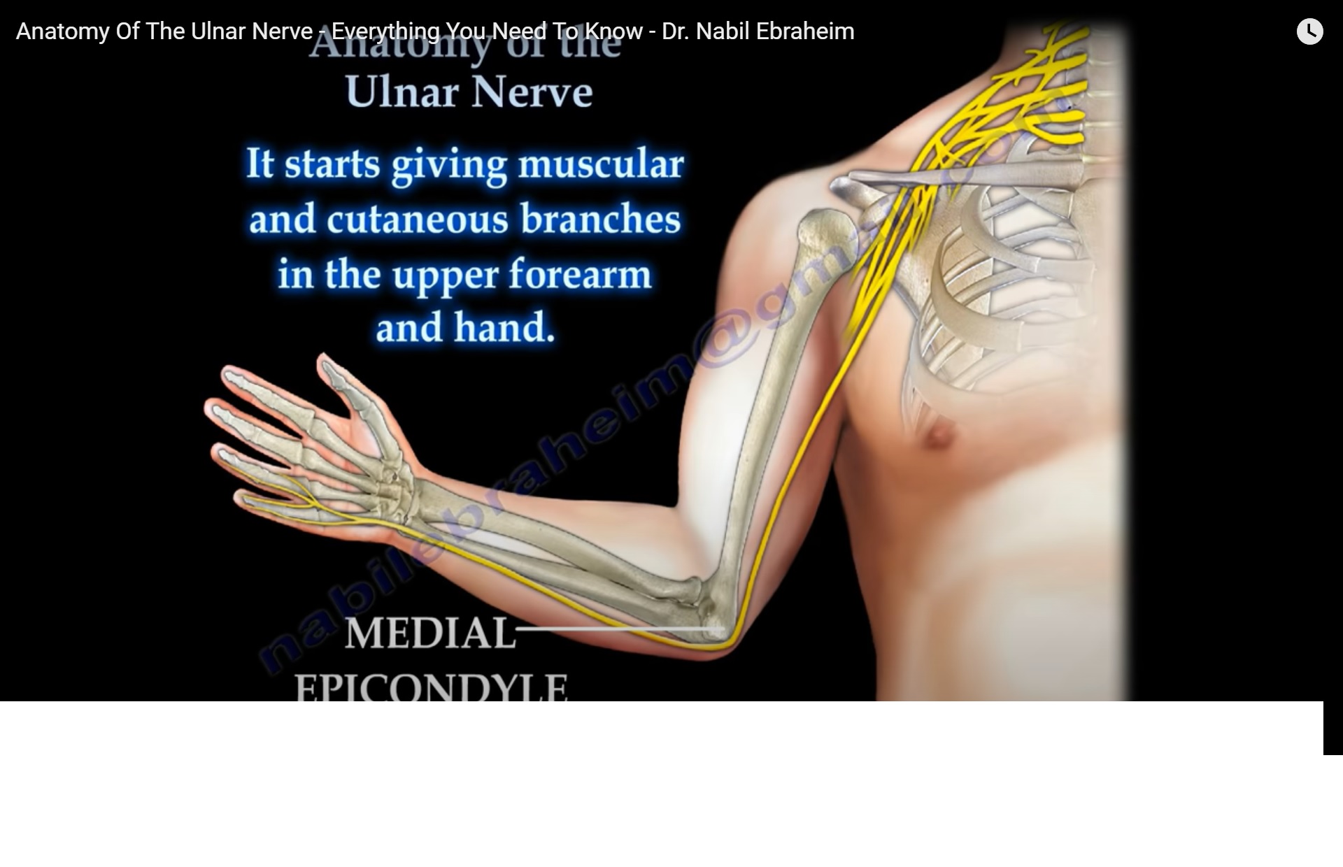
ORIGIN AND COURSE
-
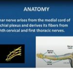
The ulnar nerve originates from the C8 and T1 nerve roots.
-
These roots form the medial cord of the brachial plexus.
-
The nerve descends along the medial aspect of the arm.
-
At the elbow, it passes posterior to the medial epicondyle of the humerus.
-
This superficial position makes the nerve vulnerable to trauma and compression.
COURSE AT THE ELBOW (CUBITAL TUNNEL)
-
Behind the medial epicondyle, the ulnar nerve runs through a fibro-osseous tunnel known as the cubital tunnel.
-
The cubital tunnel is formed by:
-
The medial epicondyle
-
The olecranon
-
The cubital tunnel retinaculum
-
-
Compression or irritation at this site results in cubital tunnel syndrome.
ENTRY INTO THE FOREARM
-
After passing behind the medial epicondyle, the ulnar nerve enters the forearm.
-
It passes between the two heads of the flexor carpi ulnaris muscle.
-
In the forearm, it travels:
-
Medial to the flexor digitorum profundus
-
Deep to the flexor carpi ulnaris
-
-
The nerve begins giving motor and sensory branches in the upper forearm.
MOTOR INNERVATION IN THE FOREARM
-
Flexor carpi ulnaris
-
Medial half of the flexor digitorum profundus (to the ring and little fingers)
COURSE AT THE WRIST
-
The ulnar nerve continues along the medial border of the forearm, adjacent to the ulna.
-
At the wrist, it lies just lateral to the pisiform bone.
-
It enters the hand through Guyon canal.
-
Within Guyon canal:
-
The ulnar nerve and ulnar artery pass superficial to the flexor retinaculum.
-
BRANCHES IN THE HAND
Deep Branch (Motor)
The deep branch of the ulnar nerve supplies:
-
Abductor digiti minimi
-
Flexor digiti minimi
-
Opponens digiti minimi
-
Third and fourth lumbricals
-
Palmar interossei
-
Dorsal interossei
-
Adductor pollicis
-
Deep head of flexor pollicis brevis
-
The ulnar nerve supplies all intrinsic hand muscles medial to the flexor pollicis longus, except the lateral two lumbricals.
Superficial Branch (Sensory)
-
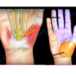
Provides palmar sensory innervation to:
-
Little finger
-
Medial half of the ring finger
-
-
Also provides dorsal sensory innervation to the same digits.
CLINICAL TEST
Tinel Sign at the Elbow
-
Performed by tapping over the ulnar nerve at the medial epicondyle.
-
A positive test produces:
-
Tingling
-
Electric shock-like sensation
-
Numbness radiating to the ring and little fingers
-
-
This sensation is similar to striking the “funny bone”.
-
Indicates ulnar nerve entrapment at the cubital tunnel.
CLINICAL FEATURES OF ULNAR NERVE COMPRESSION
-
Pain at the elbow
-
Numbness or tingling in the hand
-
Paresthesia of the fourth and fifth digits
-
Weak grip and pinch strength
-
Intrinsic muscle weakness
CLAW HAND DEFORMITY
-
Injury or entrapment of the ulnar nerve below the elbow may result in ulnar claw hand.
-
Caused by:
-
Loss of intrinsic muscle function
-
Unopposed action of the flexor digitorum profundus
-
-
Characterized by:
-
Hyperextension at the metacarpophalangeal joints
-
Flexion at the interphalangeal joints of the ring and little fingers
-
KEY POINTS
-
The ulnar nerve is most vulnerable at the medial epicondyle and Guyon canal.
-
It plays a critical role in fine motor control of the hand.
-
Early recognition of compression is essential to prevent permanent intrinsic muscle wasting.
Leave a Reply