• Skip to main content
  • Skip to secondary menu
  • Skip to primary sidebar
OrthopaedicPrinciples.com

OrthopaedicPrinciples.com

Integrating Principles and Evidence

Integrating Principles and Evidence

  • Home
  • Editorial Board
  • Our Books
    • Evidence Based Orthopaedic Principles
  • Courses
  • Exams
  • Reviews
  • Live Program
  • Contact

Triplane Fracture of Ankle

A triplane fracture of the distal tibia usually occurs during adolescence and occurs before complete closure of the distal tibial physis. The distal tibial physis (growth plate ) is a weak area which closes from central to medial, with the lateral side being the last part to close.

Triplane fracture is a Salter-Harris type IV fracture involving three planes:
1-Coronal (metaphysis) (frontal plane ) Salter type II
2-Transverse (growth plate) (horizontal plane) Salter type III
3-Sagittal (epiphysis)Salter type III

The fracture has several variations and occurs due to external rotation force. Usually occurs between 12-15 years of age.
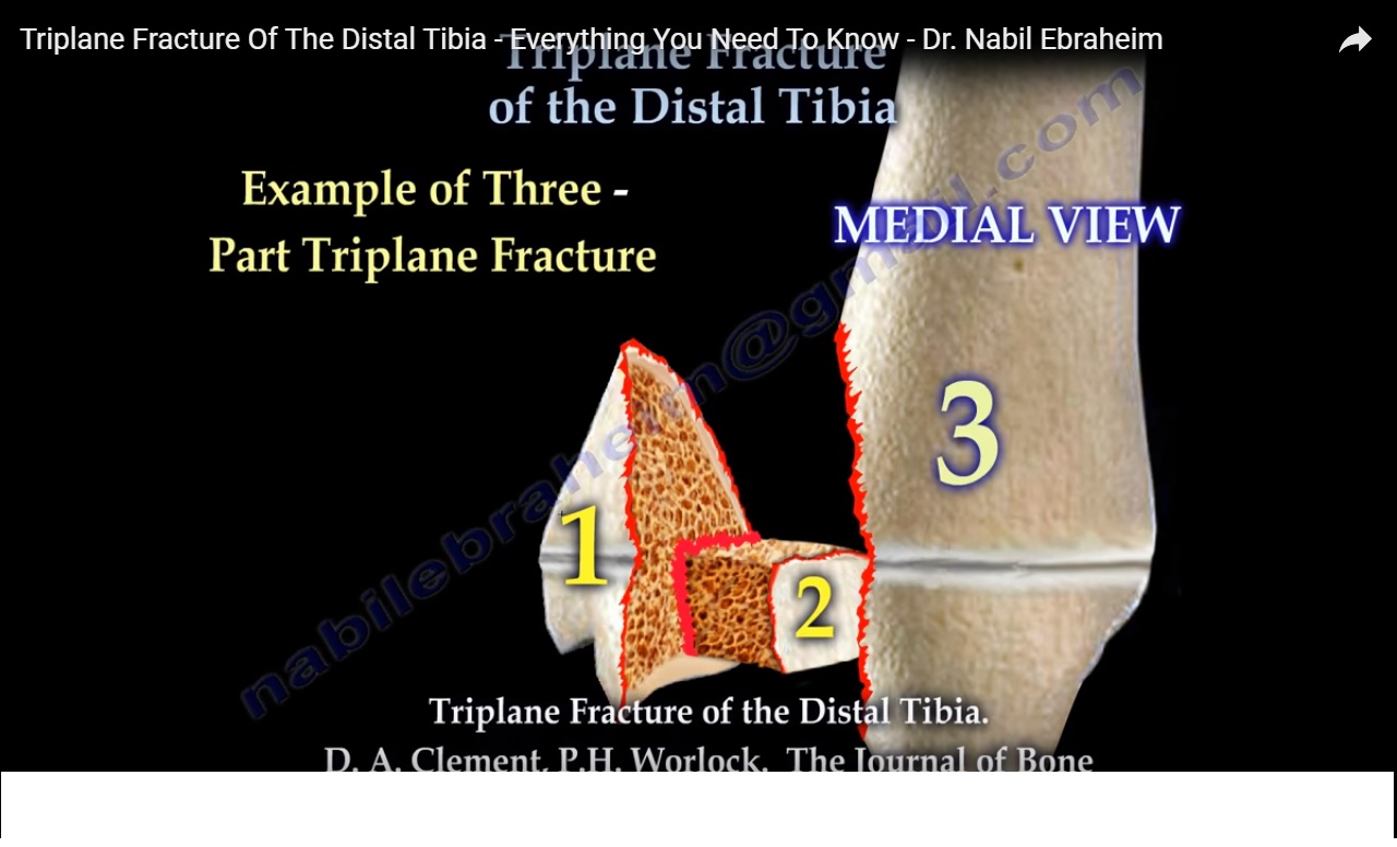
Triplane fractures are complicated three dimensional fractures. Two-part fracture is a Salter-Harris type IV. Three-part fracture is a combination of Salter-Harris type III in an AP view and type II in a lateral view.

Example of a two-fragment triplane fracture.
Example of a three-part triplane fracture. Posterior tibial metaphysis with the attached part of the posterior epiphysis. Separate anterolateral epiphyseal fragment. The anterior tibial metaphysis with the attached anteromedial part of the epiphysis.

CT scan is helpful

ORIF is the treatment if there is displacement of the fragment(s) more than 2 mm.

Post Views: 3,738

Related Posts

  • Ankle fracture in children-Tillaux Fracture

  • Posterior Malleolar Ankle Fracture

    Courtesy: Prof Nabil Ebraheim, University of Toledo, Ohio, USA

  • LisFranc Fracture

    Courtesy: Prof Nabil Ebraheim, University of Toledo, Ohio, USA

Reader Interactions

Leave a Reply

Your email address will not be published. Required fields are marked *

Primary Sidebar

Follow Us

instagram slideshare

Categories

  • -Applied Anatomy
  • -Approaches
  • -Basic Sciences
  • -Cartilage & Meniscus
  • -Classifications
  • -Examination
  • -Foot and Ankle
  • -Foot and Ankle Trauma
  • -FRCS(Tr and Orth) tutorials
  • -Gait
  • -Hand and Wrist
  • -Hand and Wrist Trauma
  • -Hand Infections
  • -Hip and Knee
  • -Hip Preservation
  • -Infections
  • -Joint Reconstruction
  • -Knee Arthroplasty
  • -Knee Preservation
  • -Metabolic Disorders
  • -Oncology
  • -OrthoBiologics
  • -OrthoPlastic
  • -Paediatric Orthopaedics
  • -Paediatric Trauma
  • -Patellofemoral Joint
  • -Pelvis
  • -Peripheral Nerves
  • -Principles
  • -Principles of Surgery
  • -Radiology
  • -Rheumatology
  • -Shoulder and Elbow
  • -Shoulder and Elbow Arthroplasty
  • -Spine Deformity
  • -Spine Oncology
  • -Spine Trauma
  • -Spine, Pelvis & Neurology
  • -Sports Ankle and Foot
  • -Sports Elbow
  • -Sports Knee
  • -Sports Medicine
  • -Sports Medicine Hip
  • -Sports Shoulder
  • -Sports Wrist
  • -Statistics
  • -Technical Tip
  • -Technology in Orth
  • -Trauma
  • -Trauma (Upper Limb)
  • -Trauma Life Support
  • -Trauma Reconstruction
  • Book Shelf
  • Book Shelf Medical
  • Careers
  • Case Studies and Free Papers
  • DNB Ortho
  • Evidence Based Orthopaedic Principles
  • Evidence Based Orthopaedics
  • Exam Corner
  • Fellowships
  • Guest Editor
  • Guest Reviews
  • Image Quiz
  • Instructional Course Lectures
  • Journal Club
  • MCQs
  • Meetings and Courses
  • Multimedia
  • News and Blog
  • Plaster Techniques
  • Podcasts
  • Public Health
  • Rehabilitation
  • Research
  • Shorts and Reels
Copyright@orthopaedicprinciples.com. All right rerserved.