• Skip to main content
  • Skip to secondary menu
  • Skip to primary sidebar
OrthopaedicPrinciples.com

OrthopaedicPrinciples.com

Integrating Principles and Evidence

Integrating Principles and Evidence

  • Home
  • Editorial Board
  • Our Books
    • Evidence Based Orthopaedic Principles
  • Courses
  • Exams
  • Reviews
  • Live Program
  • Contact

Tibialis Anterior Muscle Anatomy


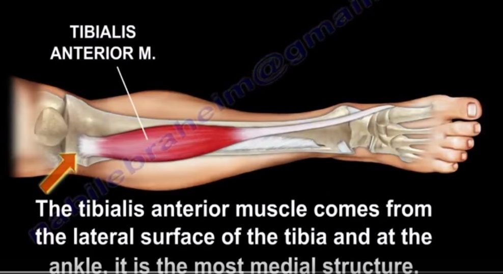
ANATOMY OF TIBIALIS ANTERIOR MUSCLE

The leg has four muscle compartments

  • Anterior
  • Lateral
  • Superficial posterior
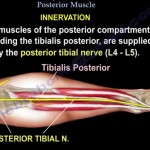
  • Deep posterior

Anterior compartment

  • Nerve within the compartment- Deep peroneal nerve
  • Muscles include : Tibialis anterior, extensor hallucis longus and extensor digitorum longus

Origin

  • Upper half and lateral condyle of tibia

Insertion

  • Medial cuneiform and first metatarsal bone of foot

Course

  • The tendon of tibialis anterior along with other extensor tendons pass under the extensor retinaculum

Structures of anterior compartment

  • Tibialis anterior
  • Flexor hallucis longus
  • Extensor digitorum longus
  • Peronius tertius
  • Anterior tibial artery
  • Deep peroneal nerve

Nerve supply of tibialis anterior muscle: Deep peroneal nerve

INTERRUPTION OF NERVE SUPPLY

  • Pelvic fracture affecting L4-L5 root
  • Posterolateral disc herniationL3,L4
  • Foraminal disc herniationL4-L5
  • Sciatic nerve injury

FUNCTION OF TIBIALIS ANTERIOR

  • Dorsal flexion of ankle
  • Invertion of hind foot
  • Tibialis anterior dorsiflexes the foot in preparation for heel strike during the last swing phase of the gait and it eccentrically contracts after heel strike to slow progression to foot flat.

RUPTURE OF TIBIALIS ANTERIOR TENDON

  • Cause loss of dorsiflexion of ankle
  • May occur due to laceration
  • May also be a closed rupture

Closed rupture

  • It occurs due to strong eccentric contracture in young patients or attenuation rupture in elderly who have diabetes , inflammatory arthritis or from steroid injections.

Presentation

• Difficulty clearing the foot during the gait with chronic rupture
• In acute rupture, will hear a POP and there will be swelling of the anterior ankle

1. Foot drop
2. Steppage gait
3. Pseudo tumour
4. Anterior ankle pain
5. Weak dorsiflexion
6. No tendon is felt in resisted dorsiflexion

TREATMENT

• PARTIAL TEAR: CAST APPLICATION

• COMPLETE TEAR: AFO in low demand patients , however surgical repair proves to be successful in elderly
SURGERY

BEST CHOICE
• Acute repair , end to end repair when the tendon is normal
• In chronic rupture do autograft (EHL) or allograft


Important points

• When foot drop occurs in compartment syndrome it is due to tibialis anterior

Charot-Marie-Tooth disease
(peroneal muscular atrophy) the affected muscles become weak:

– Tibialis Anterior

– Peroneus Brevis

– Intrinsic muscles of the hand and foot

The weak tibialis anterior muscle is overpowered by the unaffected peroneus longus muscle, bringing
the first ray into a plantarflexed position.

3 tibialis anterior J

Courtesy: Prof Nabile Ebraheim, University of Toledo, Ohio, USA

Post Views: 6,616

Related Posts

  • Tibialis Posterior Anatomy

    Courtesy: Prof Nabile Ebraheim, University of Toledo, Ohio, USA Anatomy of the Tibialis Posterior Muscle…

  • Popliteus Muscle

    Courtesy : Prof Nabile Ebraheim, University of Toledo, Ohio, USA Popliteus Muscle A triangular muscle…

  • Muscle Anatomy of the Trunk

    Courtesy: Prof Nabil Ebraheim, University of Toledo, Ohio, USA

Reader Interactions

Leave a Reply

Your email address will not be published. Required fields are marked *

Primary Sidebar

Follow Us

instagram slideshare

Categories

  • -Applied Anatomy
  • -Approaches
  • -Basic Sciences
  • -Cartilage & Meniscus
  • -Classifications
  • -Examination
  • -Foot and Ankle
  • -Foot and Ankle Trauma
  • -FRCS(Tr and Orth) tutorials
  • -Gait
  • -Hand and Wrist
  • -Hand and Wrist Trauma
  • -Hand Infections
  • -Hip and Knee
  • -Hip Preservation
  • -Infections
  • -Joint Reconstruction
  • -Knee Arthroplasty
  • -Knee Preservation
  • -Metabolic Disorders
  • -Oncology
  • -OrthoBiologics
  • -OrthoPlastic
  • -Paediatric Orthopaedics
  • -Paediatric Trauma
  • -Patellofemoral Joint
  • -Pelvis
  • -Peripheral Nerves
  • -Principles
  • -Principles of Surgery
  • -Radiology
  • -Rheumatology
  • -Shoulder and Elbow
  • -Shoulder and Elbow Arthroplasty
  • -Spine Deformity
  • -Spine Oncology
  • -Spine Trauma
  • -Spine, Pelvis & Neurology
  • -Sports Ankle and Foot
  • -Sports Elbow
  • -Sports Knee
  • -Sports Medicine
  • -Sports Medicine Hip
  • -Sports Shoulder
  • -Sports Wrist
  • -Statistics
  • -Technical Tip
  • -Technology in Orth
  • -Trauma
  • -Trauma (Upper Limb)
  • -Trauma Life Support
  • -Trauma Reconstruction
  • Book Shelf
  • Book Shelf Medical
  • Careers
  • Case Studies and Free Papers
  • DNB Ortho
  • Evidence Based Orthopaedic Principles
  • Evidence Based Orthopaedics
  • Exam Corner
  • Fellowships
  • Guest Editor
  • Guest Reviews
  • Image Quiz
  • Instructional Course Lectures
  • Journal Club
  • MCQs
  • Meetings and Courses
  • Multimedia
  • News and Blog
  • Plaster Techniques
  • Podcasts
  • Public Health
  • Rehabilitation
  • Research
  • Shorts and Reels
Copyright@orthopaedicprinciples.com. All right rerserved.