Courtesy: Prof Nabil Ebraheim, University of Toledo, Ohio, USA
LATERAL ROTATORS OF THE HIP
There are six short external rotators of the hip:
-
Piriformis
-
Superior gemellus
-
Obturator internus
-
Inferior gemellus
-
Obturator externus
-
Quadratus femoris
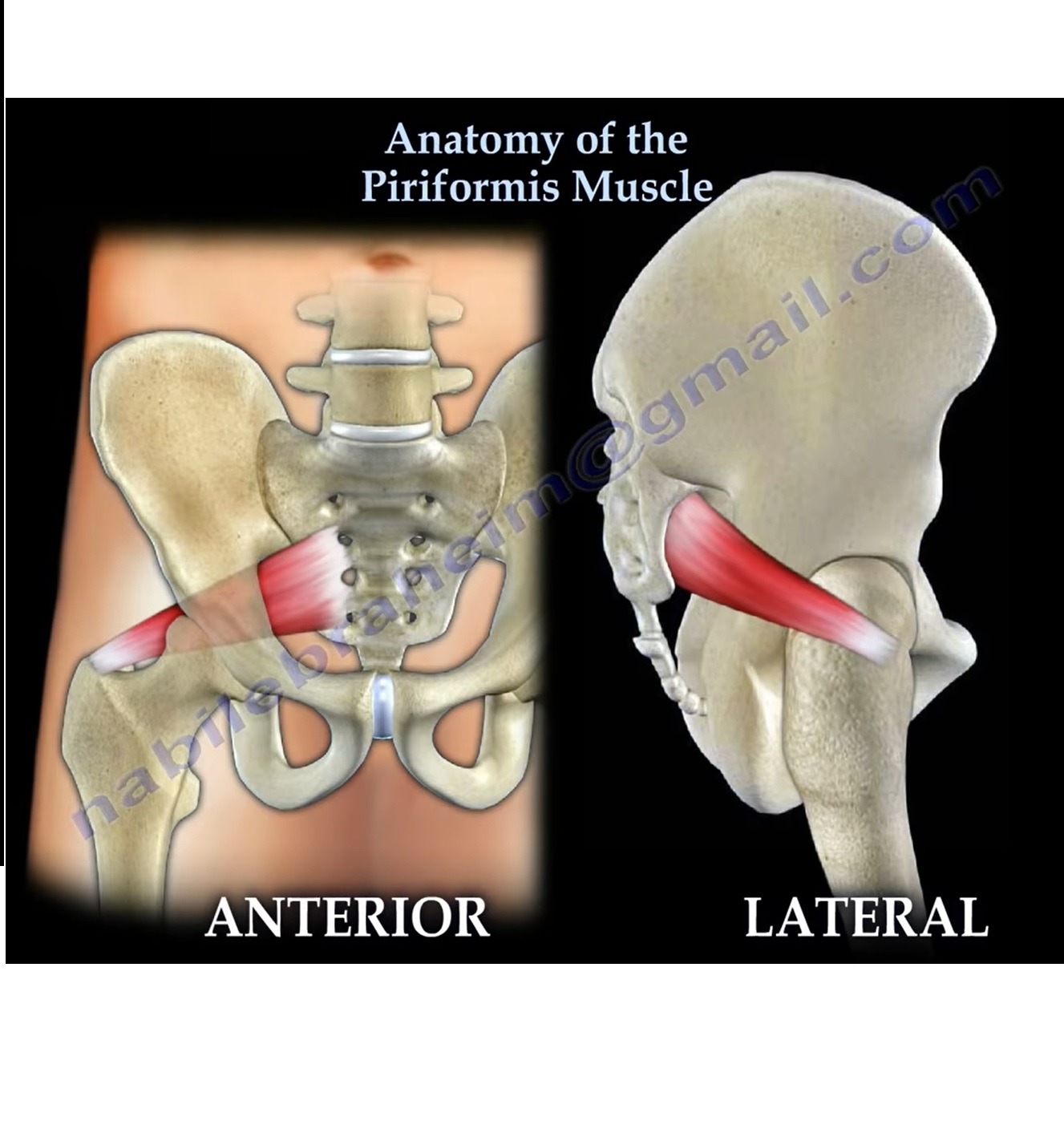
ORIGIN
-
Arises from:
-
The anterior surface of the lateral process of the sacrum
-
The capsule of the sacroiliac joint
-
The gluteal surface of the ilium near the margin of the greater sciatic notch
-
INSERTION
-
Inserts into the superior border of the greater trochanter of the femur
RELATIONS (POSTERIOR)
Structures related posteriorly to the piriformis muscle include:
-
Sciatic nerve (normally passes inferior to the muscle)
-
Superior gluteal artery
-
Superior gluteal nerve
-
Nerve to piriformis (nerve roots L5, S1, S2)
ACTIONS
-
Lateral rotation of the hip
-
Abduction of the hip when the hip is flexed
ANATOMICAL VARIATIONS OF THE SCIATIC NERVE
-
The sciatic nerve is composed of:
-
Tibial division
-
Common peroneal division
-
-
These divisions are usually bound together but may separate in relation to the piriformis muscle.
Variations in Relationship
-
Sciatic nerve passes beneath an undivided piriformis muscle (most common).
-
Piriformis muscle is divided, with the common peroneal division passing between the two heads.
-
Common peroneal division passes over the piriformis, while the tibial division passes beneath.
-
Entire sciatic nerve passes through a divided piriformis muscle.
CLINICAL SIGNIFICANCE
Piriformis Fossa
-
The piriformis fossa is the proximal entry point for most intramedullary femoral nail insertions.
PIRIFORMIS SYNDROME
Definition
-
A condition in which the sciatic nerve is compressed by the piriformis muscle or associated fibrous bands.
Diagnosis
-
Requires a high index of clinical suspicion
-
Based primarily on:
-
Patient history
-
Physical examination
-
-
Electromyography and bone scan are generally not helpful
Symptoms
-
May follow blunt trauma to the buttock
-
Localized buttock pain
-
Pain aggravated by:
-
Sitting
-
Driving
-
Bicycling
-
Running, especially in young individuals
-
-
Tenderness in the region of the sciatic notch
-
Radicular pain and paresthesia are uncommon
Clinical Signs
-
Deep palpation over the greater sciatic notch reproduces pain
Provocative Test
Lasègue Maneuver
-
Hip flexed to 90 degrees with knee extension
-
Reproduction of pain indicates compression of the sciatic nerve
-
Compression may be due to:
-
Piriformis muscle
-
Fibrous bands
-
Vascular anomalies
-
Imaging
Magnetic Resonance Imaging Findings
-
Enlarged piriformis muscle
-
Vascular anomalies
-
Compression or displacement of the sciatic nerve
TREATMENT
Conservative Management
-
Aquatic therapy
-
Physiotherapy
-
Nonsteroidal anti-inflammatory medications
-
Local injections
Surgical Management
-
Reserved as a last resort
-
Involves:
-
Surgical release of the piriformis muscle
-
Decompression of the sciatic nerve
-
KEY POINTS
-
The piriformis muscle is a critical anatomical landmark in the deep gluteal region.
-
Variations in sciatic nerve anatomy explain differing clinical presentations.
-
Most cases of piriformis syndrome respond to conservative treatment.
-
Accurate anatomical knowledge is essential during hip surgery and intramedullary nailing.


This is an very important muscle and preservation of this muscle has been known to help in reducing posterior dislocations of the Hip Joint.
References:
1.. Piriformis and obturator internus morphology: a cadaveric study. Clinical Anatomy 01/2011
24(1):70-6.
2.. INCIDENCE OF PIRIFORMIS TENDON PRESERVATION ON THE DISLOCATION RATE
OF TOTAL HIP REPLACEMENT FOLLOWING THE POSTERIOR APPROACH,
by Charbel D. MOUSSALLEM,Fadi A. HOYEK and Jean-Claude F. LAHOUD
in the Lebanese Medical Journal 2012 – Volume 60 (1) 19
Dr.K.Mohan Iyer,Senior Consultant Orthopedic Surgeon,Bangalore,India