Courtesy: Prof Nabil Ebraheim, University of Toledo, Ohio, USA
Tennis Elbow (Lateral Epicondylitis)
Definition
-
Tennis elbow, also called lateral epicondylitis, is an overuse injury.
-
It causes:
-
Inflammation
-
Tendinosis
-
Lateral elbow pain
-
-
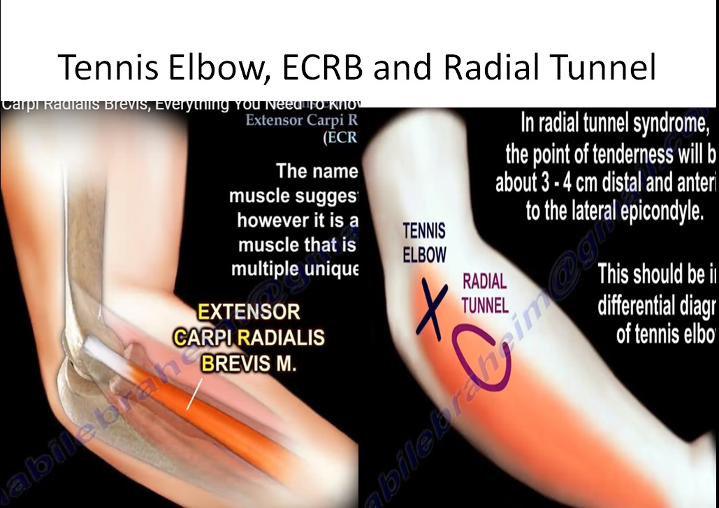
The pathology occurs at the origin of the Extensor Carpi Radialis Brevis (ECRB) muscle.
-
Pain is located on the outer (lateral) side of the elbow.
-
Symptoms may interfere with:
-
Sleep
-
Daily activities
-
Carrying groceries
-
Etiology and Risk Factors
-
It is the most common cause of elbow pain.
-
Approximately 50% of tennis players develop tennis elbow.
-
Contributing factors in tennis players:
-
Incorrect grip size
-
Poor swing technique
-
-
Also affects workers performing:
-
Heavy lifting
-
Repetitive gripping
-
Use of heavy tools
-
Pathology
-
The condition usually begins with microtears at the origin of the Extensor Carpi Radialis Brevis.
-
Caused by eccentric overload.
-
Aggravated by:
-
Repetitive wrist extension
-
Forearm pronation
-
-
Tendon pathology shows:
-
Disorganized collagen
-
Angiofibroblastic hyperplasia
-
Vascular hyperplasia
-
Fibroblast hypertrophy
-
Replacement of tendon fibers with vascular granulation tissue
-
Clinical Features
-
Lateral-sided elbow pain
-
Pain with gripping
-
Decreased grip strength
-
Pain with repetitive wrist extension
-
Tenderness around the lateral epicondyle
-
Point tenderness on palpation over the lateral epicondyle
Physical Examination
-
Diagnosis is based on symptoms and physical examination.
-
Provocative test:
-
Resisted wrist extension
-
Elbow fully extended
-
Reproduction of pain at the lateral epicondyle
-
Differential Diagnosis
Radial Tunnel Syndrome
-
Occurs in approximately 5% of patients.
-
Caused by compression of the posterior interosseous nerve.
-
Pain is located:
-
3–4 cm distal and anterior to the lateral epicondyle.
-
-
Should be suspected if the patient does not recover as expected with tennis elbow treatment.
-
Diagnostic approach:
-
Injection into the radial tunnel to assess response.
-
Other Conditions to Rule Out
-
Cervical disc pathology
-
Triceps tendinitis
Investigations
-
Diagnosis is primarily clinical.
-
X-rays are usually normal.
-
Rarely, calcification may be seen.
Treatment
Nonoperative Treatment
-
Activity modification
-
Nonsteroidal anti-inflammatory medication
-
Ice
-
Physiotherapy:
-
Tendon gliding exercises
-
Eccentric conditioning
-
Strengthening program
-
-
Bracing
-
Up to 95% success rate with nonoperative treatment.
-
Improvement may take 6–12 months.
-
Condition often improves with time regardless of the conservative method chosen.
Injection Therapy
-
Steroid injection (up to three injections)
-
Useful for controlling acute symptoms.
-
-
Platelet-Rich Plasma (PRP) injection
-
Ultrasound guidance may be helpful during injection.
-
Physiotherapy may provide better symptomatic relief at one year compared to steroid injection.
Surgical Treatment
-
Considered the last resort.
-
Procedure involves:
-
Release and debridement of the Extensor Carpi Radialis Brevis origin.
-
-
Excessive debridement or release may injure the lateral collateral ligament.
-
This may lead to posterolateral rotatory instability of the elbow.
Extensor Carpi Radialis Brevis (ECRB) Muscle
Origin
-
Lateral epicondyle of the humerus
-
Part of the common extensor tendon
Insertion
-
Base of the dorsal aspect of the third metacarpal
Innervation
-
Radial nerve
Function
-
Wrist extension
-
Abduction of the hand at the wrist joint
Location
-
Located in the second dorsal compartment on the radial side of Lister’s tubercle.
Other Conditions Involving the ECRB Muscle
1. Intersection Syndrome
-
Inflammation at the crossing of:
-
First dorsal compartment
-
Second dorsal compartment
-
-
The second dorsal compartment contains the Extensor Carpi Radialis Brevis.
-
Caused by repetitive wrist extension.
-
Tenderness is present on the dorsoradial aspect of the forearm.
-
Approximately 5 cm proximal to the wrist joint.
-
Involves intersection of:
-
Extensor Carpi Radialis Longus
-
Extensor Carpi Radialis Brevis
-
Abductor Pollicis Longus
-
Extensor Pollicis Brevis
-
2. Posterolateral Elbow Instability
-
May occur after excessive release of the Extensor Carpi Radialis Brevis tendon during tennis elbow surgery.
-
Due to proximity of the ECRB origin to the ulnar humeral (lateral ulnar collateral) ligament.
-
Injury to this ligament may cause posterolateral elbow instability.
3. Tendon Transfer in High Radial Nerve Palsy
-
High radial nerve palsy results in wrist drop.
-
Caused by paralysis of the extensor tendons.
-
Surgical management:
-
Pronator teres is transferred to the Extensor Carpi Radialis Brevis.
-
-
Purpose:
-
Restore wrist extension
-

Leave a Reply