
Courtesy: Dr Thiago Busato MD, Hip Surgeon, Curitiba, Brazil
Surgical Hip Dislocation: Principles, Technique and Current Indications
Introduction
- Surgical hip dislocation is a specialized hip-preservation approach that provides extensive exposure of the femoral head and acetabulum while aiming to preserve the blood supply to the femoral head.
- The technique was developed and popularized by Reinhold Ganz and colleagues based on detailed anatomical studies of the vascularity of the proximal femur.
- The modern technique combines:
- A lateral position
- Trochanteric flip osteotomy
- Preservation of the short external rotators
- Protection of the medial femoral circumflex artery
- Controlled anterior dislocation of the femoral head
- The principal advantage is the ability to directly visualize and dynamically assess both the femoral head and acetabulum.
- Surgical hip dislocation is now primarily reserved for complex hip deformities and selected traumatic conditions, because less invasive procedures are available for many routine cases.
Historical Background
- The concept of femoroacetabular impingement is not new.
- Early descriptions recognized mechanical conflict between the femoral neck and acetabular rim as a cause of hip pain and secondary degenerative changes.
- Historically, hip preservation included procedures such as:
- Varus osteotomy
- Valgus osteotomy
- Proximal femoral osteotomy
- The development of surgical hip dislocation represented a major change because the surgeon could directly address the underlying intra-articular and extra-articular deformity.
- The technique subsequently became an important tool in the development of modern hip-preservation surgery.
Anatomical Basis
Blood Supply of the Femoral Head
The safety of surgical hip dislocation depends principally on understanding the vascular anatomy of the proximal femur.
Medial Femoral Circumflex Artery
- The medial femoral circumflex artery provides the dominant blood supply to the adult femoral head.
- Its deep branch gives rise to superior retinacular vessels that course along the femoral neck.
- These vessels are particularly important for maintaining femoral-head perfusion.
- The technique is designed to preserve these vessels.
Structures Protecting the Blood Supply
- The obturator externus and short external rotator structures contribute to protection of the vascular pedicle.
- The posterior approach and trochanteric flip technique avoid unnecessary disruption of these structures.
- The short external rotators are therefore not routinely detached.
The original anatomical and clinical description reported no cases of avascular necrosis in 213 hips using the technique, although modern clinical experience demonstrates that femoral-head osteonecrosis cannot be considered impossible.
Principles of Surgical Hip Dislocation
The essential principles are:
- Understand the vascular anatomy.
- Preserve the medial femoral circumflex artery and its retinacular branches.
- Perform a controlled trochanteric flip osteotomy.
- Preserve the short external rotators.
- Perform capsulotomy carefully.
- Dislocate the femoral head in a controlled manner.
- Avoid excessive traction or twisting of the femoral neck.
- Perform the required femoral and acetabular procedures under direct visualization.
- Reduce the hip gently.
- Fix the greater trochanter securely.
Advantages
Extensive Exposure
Surgical hip dislocation provides:
- Near circumferential visualization of the femoral head
- Direct visualization of the acetabulum
- Access to the femoral head-neck junction
- Access to the acetabular rim
- Assessment of cartilage
- Assessment of the labrum
- Dynamic assessment of femoroacetabular conflict
Dynamic Assessment
One major advantage is the ability to assess the hip through a range of motion while directly observing:
- Femoral head-neck impingement
- Acetabular rim conflict
- Cam morphology
- Pincer morphology
- Cartilage lesions
- Labral pathology
Treatment of Complex Deformity
Unlike procedures designed for relatively straightforward femoroacetabular impingement, surgical dislocation can be useful when there is:
- Severe proximal femoral deformity
- Complex femoral head-neck morphology
- Post-traumatic deformity
- Deformity following slipped capital femoral epiphysis
- Legg-Calvé-Perthes disease sequelae
- Complex femoral head pathology
Limitations
Despite its excellent exposure, surgical hip dislocation has important disadvantages:
- Greater soft-tissue dissection than hip arthroscopy
- Greater trochanteric osteotomy
- Risk of trochanteric nonunion or fixation failure
- Heterotopic ossification
- Postoperative abductor weakness
- Longer rehabilitation
- Greater postoperative morbidity than minimally invasive techniques
- Potential neurovascular injury
- Risk of femoral-head osteonecrosis if vascular principles are violated
Systematic reviews have reported good clinical improvement but also higher reoperation or complication rates with open surgical dislocation compared with less invasive approaches in selected femoroacetabular impingement populations.
Patient Selection
Appropriate patient selection is critical.
Favourable Features
- Symptomatic patient
- Young or relatively young adult
- Minimal or absent established osteoarthritis
- Preserved cartilage
- Correctable structural deformity
- Patient understands the rehabilitation requirements
- Failure of appropriate nonoperative treatment where indicated
Unfavourable Features
- Advanced osteoarthritis
- Extensive irreversible cartilage damage
- Severe joint-space loss
- Poor biological potential
- Significant medical comorbidity
- Inability to comply with postoperative restrictions
Important Point
Age alone should not be used as an absolute cutoff.
The decision to perform hip-preservation surgery should depend on:
Symptoms + joint preservation potential + cartilage status + deformity + patient goals + surgeon experience.
Advanced cartilage damage and established osteoarthritis are stronger negative prognostic factors than chronological age alone.
Preoperative Evaluation
Clinical Assessment
Assess:
- Pain location
- Mechanical symptoms
- Range of motion
- Impingement signs
- Gait
- Abductor function
- Limb-length discrepancy
- Previous trauma or surgery
- Functional limitations
Radiographs
Useful radiographic assessment includes:
- Anteroposterior pelvis
- Dunn or other lateral views
- Frog-leg lateral view where appropriate
- False-profile view when acetabular coverage requires assessment
Evaluate:
- Cam morphology
- Acetabular coverage
- Acetabular version
- Joint space
- Osteoarthritis
- Proximal femoral deformity
- Greater trochanter position
- Coxa vara or coxa valga
Computed Tomography
Computed tomography, particularly three-dimensional reconstruction, may be valuable in:
- Complex proximal femoral deformity
- Post-traumatic deformity
- Slipped capital femoral epiphysis sequelae
- Legg-Calvé-Perthes disease sequelae
- Surgical planning
- Assessment of complex femoral head or acetabular morphology
Magnetic Resonance Imaging
Magnetic resonance imaging can assess:
- Labrum
- Articular cartilage
- Subchondral bone
- Femoral-head viability
- Muscle quality
- Associated soft-tissue pathology
Surgical Technique
- Patient Position
- The patient is placed in the lateral decubitus position.
- The operative limb should be freely mobile.
- Adequate padding is essential.
- The limb should be positioned so that the hip can be flexed, abducted and externally rotated during the procedure.
- Surgical Approach
- A modified posterior or Gibson-type approach is commonly used.
- The incision is centred over the greater trochanter.
- The fascia lata is opened.
- The greater trochanter and gluteal structures are identified.
- The trochanteric flip osteotomy is then planned.
- Trochanteric Flip Osteotomy
- A thin fragment of the greater trochanter is osteotomized.
- The osteotomy extends sufficiently to maintain a robust trochanteric fragment while allowing it to be mobilized anteriorly.
- A stepped osteotomy may provide greater stability during fixation.
- The fragment should contain the necessary abductor attachment.
- The vastus lateralis attachment is managed carefully.
Important Principle
The osteotomy should preserve adequate bone stock to minimize:
- Trochanteric fracture
- Fixation failure
- Nonunion
- Exposure of the Capsule
- The trochanteric fragment is mobilized.
- The interval between the external rotators and gluteal musculature is developed.
- The posterior capsule is exposed.
- The hip is positioned appropriately to facilitate anterior dislocation.
- Capsulotomy
A Z-shaped capsulotomy can be used.
The capsulotomy permits:
- Adequate visualization
- Controlled dislocation
- Preservation of capsular tissue for subsequent closure
Capsular preservation and repair are important components of contemporary hip-preservation surgery.
- Hip Dislocation
- The hip is carefully dislocated anteriorly.
- A bone hook may assist controlled dislocation.
- The operative limb is supported throughout the maneuver.
- The femoral head should not be forcibly levered against the acetabular rim.
Critical Principle
The femoral head should be dislocated only after the vascular structures have been identified and protected.
- Assessment of Femoral Head Perfusion
- Intraoperative assessment of femoral-head vascularity has historically been described.
- A small drill hole in the femoral head can be used in some techniques to assess bleeding.
- Persistent bleeding from cancellous bone provides reassurance regarding femoral-head perfusion.
This assessment should be regarded as an adjunct rather than a substitute for meticulous preservation of the vascular anatomy.
- Treatment of Femoroacetabular Impingement
Once the hip is dislocated, the entire femoral head-neck junction can be inspected.
Possible procedures include:
- Cam resection
- Femoral head-neck reshaping
- Acetabular rim trimming
- Labral repair or reconstruction
- Cartilage procedures
- Removal of intra-articular loose bodies
- Treatment of complex deformity
The major advantage is direct visualization of the deformity and the ability to assess the correction dynamically.
Complex Hip Deformities
Surgical hip dislocation is particularly useful when conventional arthroscopy cannot adequately address the deformity.
Examples include:
Slipped Capital Femoral Epiphysis Sequelae
Possible abnormalities include:
- Severe metaphyseal prominence
- Cam morphology
- Reduced hip flexion
- Femoroacetabular impingement
- Secondary cartilage damage
Selected severe deformities may require open corrective procedures rather than isolated arthroscopy. Surgical dislocation has been used in complex cases, although complication rates may be higher than with arthroscopy in less severe deformities.
Legg-Calvé-Perthes Disease Sequelae
Possible deformities include:
- Coxa magna
- Coxa breva
- Femoral-head enlargement
- Aspherical femoral head
- High-riding greater trochanter
- Trochanteric overgrowth
- Functional abductor insufficiency
- Acetabular dysplasia or borderline coverage
- Secondary impingement
Surgical dislocation provides direct access for selected reconstructive procedures.
Possible procedures include:
- Femoral head-neck reshaping
- Relative femoral neck lengthening
- Trochanteric advancement
- Labral treatment
- Cartilage procedures
- Correction of impingement
Femoral Head Reduction Osteotomy
- This is a highly specialized reconstructive procedure.
- It may be considered in selected patients with severe femoral-head deformity, particularly when preservation of the native hip is preferable to immediate arthroplasty.
- The objective is to reconstruct a more spherical, congruent femoral head.
- It is technically demanding and requires careful assessment of:
- Cartilage
- Femoral-head vascularity
- Remaining viable bone
- Acetabular congruence
- Patient expectations
It should not be presented as a routine procedure or as a guaranteed method of avoiding eventual total hip arthroplasty.
Traumatic Indications
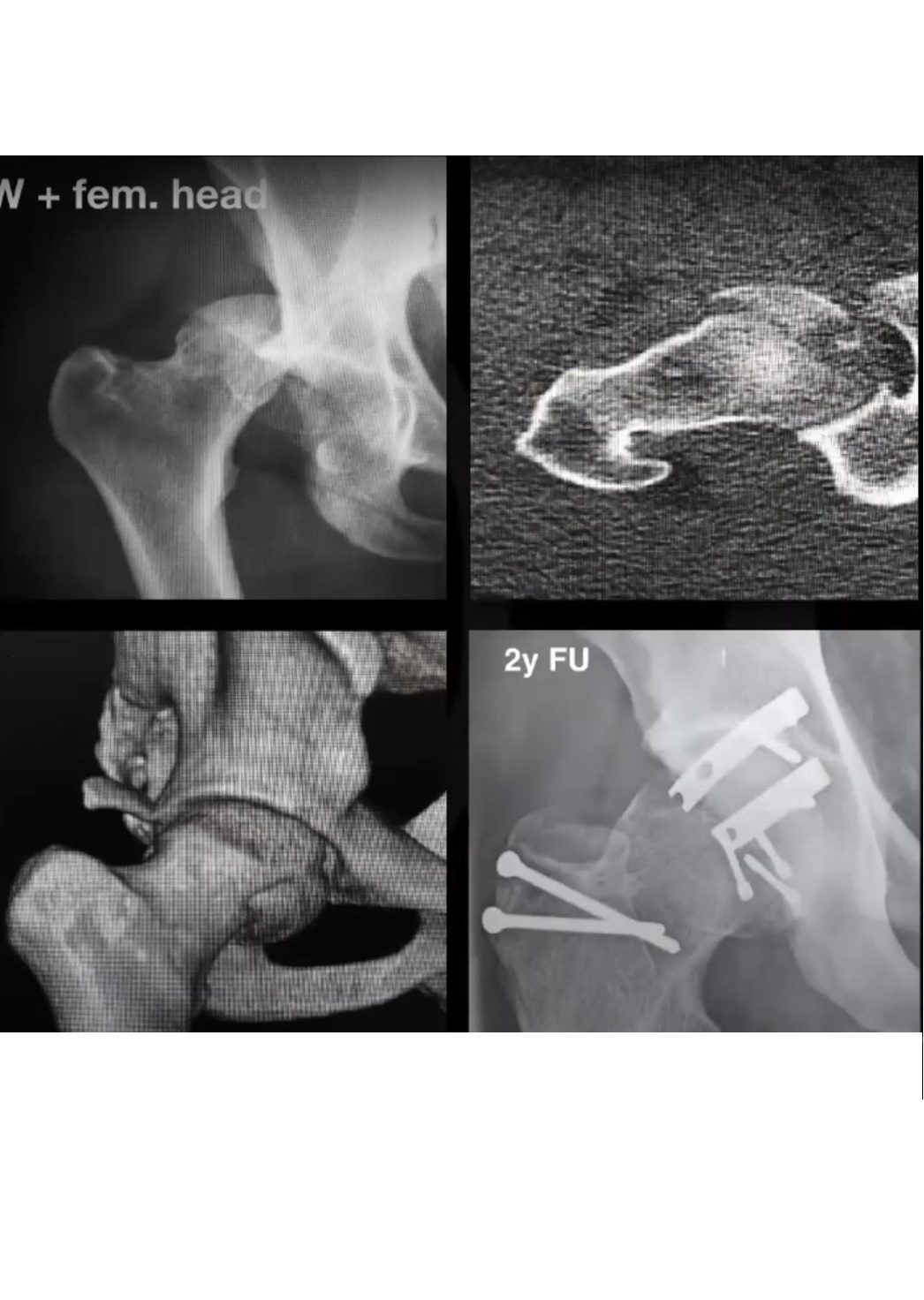
Surgical hip dislocation has an important role in selected complex hip trauma.
Femoral Head Fractures
It may be considered for:
- Selected Pipkin fractures
- Complex femoral-head fractures
- Femoral-head fracture associated with posterior wall fracture
- Irreducible fracture-dislocations
- Fractures requiring direct visualization for accurate reduction
Acetabular Fractures
Selected cases may include:
- Complex posterior wall fractures
- Posterior wall plus posterior column fractures
- Femoral head and acetabular fractures
- Transverse fracture patterns where direct visualization is advantageous
The approach allows simultaneous assessment and treatment of:
- Femoral head
- Acetabulum
- Labrum
- Articular cartilage
However, the approach should not automatically replace anterior or posterior approaches. Fracture morphology, fragment location and vascular considerations determine the appropriate approach.
Advantages in Femoral Head Fractures
Potential benefits include:
- Direct visualization of the entire femoral head
- Accurate fracture reduction
- Direct assessment of cartilage
- Ability to identify associated labral injury
- Ability to address selected acetabular injuries through the same exposure
- Ability to assess associated soft-tissue injuries
Alternative approaches may be preferable depending on fracture orientation and associated acetabular involvement.
Fixation of the Greater Trochanter
- After completion of the intra-articular procedure, the hip is reduced.
- The trochanteric fragment is repositioned anatomically.
- Fixation may be performed with:
- Screws
- Specialized trochanteric fixation constructs
- Stable fixation is essential to permit healing.
Postoperative Rehabilitation
A typical protocol includes:
- Protected weight bearing during the early healing period
- Crutches as required
- Gradual restoration of hip range of motion
- Progressive strengthening
- Particular attention to abductor rehabilitation
The exact duration of protected weight bearing should depend on:
- Osteotomy configuration
- Fixation stability
- Bone quality
- Associated procedures
- Radiographic evidence of healing
The commonly used approach of approximately 6 weeks of protected weight bearing is a protocol rather than a universal rule.
Complications
- Greater Trochanteric Complications
- Nonunion
- Delayed union
- Displacement
- Fixation failure
- Symptomatic hardware
- Abductor weakness
These are among the characteristic complications of surgical hip dislocation.
- Heterotopic Ossification
- Heterotopic ossification may occur after open hip surgery.
- Prophylaxis may be considered according to patient risk and institutional protocol.
- Nonsteroidal anti-inflammatory drugs are commonly used when not contraindicated.
- Femoral-Head Osteonecrosis
- The major feared complication is disruption of femoral-head vascularity.
- Risk is minimized by:
- Preserving the medial femoral circumflex artery
- Preserving the obturator externus
- Avoiding unnecessary soft-tissue stripping
- Using a controlled dislocation technique
The original Ganz series demonstrated the vascular safety of the technique when performed correctly, but subsequent literature confirms that osteonecrosis remains a potential complication rather than an impossible event.
- Nerve Injury
Potential injuries include:
- Sciatic nerve injury
- Femoral nerve injury
- Lateral femoral cutaneous nerve injury
- Infection
- Superficial or deep infection may occur.
- Persistent Pain or Failure
Possible causes include:
- Incomplete correction
- Advanced cartilage damage
- Incorrect diagnosis
- Residual impingement
- Progression of osteoarthritis
- Poor patient selection
Surgical Hip Dislocation Versus Hip Arthroscopy
| Feature | Surgical hip dislocation | Hip arthroscopy |
| Exposure | Extensive | Limited but expanding |
| Femoral head visualization | Excellent | Limited |
| Acetabular visualization | Excellent | Excellent within accessible regions |
| Dynamic assessment | Excellent | Possible but more limited |
| Complex deformity | Particularly useful | Best for selected deformities |
| Cam resection | Excellent | Excellent in appropriate cases |
| Greater trochanteric osteotomy | Required | Not required |
| Soft-tissue morbidity | Greater | Lower |
| Rehabilitation | Longer | Usually shorter |
| Trochanteric complications | Possible | Avoided |
| Routine uncomplicated FAI | Generally not preferred | Commonly preferred |
| Complex trauma | Useful in selected cases | Limited for some patterns |
| Severe deformity | Major role | May be insufficient |
Modern evidence suggests that arthroscopy is generally preferable for appropriately selected straightforward femoroacetabular impingement, whereas surgical dislocation retains an important role for complex morphology and selected trauma.
Current Indications
The contemporary role of surgical hip dislocation includes:
Hip Preservation
- Complex femoroacetabular impingement
- Severe cam deformity
- Complex post-slipped-capital-femoral-epiphysis deformity
- Selected Legg-Calvé-Perthes sequelae
- Complex femoral-head deformity
- Selected rotational abnormalities
- Femoral-head reconstruction procedures
Trauma
- Selected femoral-head fractures
- Complex fracture-dislocations
- Selected posterior wall acetabular fractures
- Femoral-head and acetabular fracture combinations
- Irreducible traumatic hip dislocation
- Selected cases requiring direct intra-articular visualization
The literature over the last 2 decades shows that femoroacetabular impingement remains the most common non-traumatic indication, while femoral-head fractures are an important traumatic indication.
Contraindications and Poor Prognostic Factors
Relative or Strong Contraindications
- Advanced osteoarthritis
- Severe cartilage loss
- Poor-quality remaining joint
- Inability to comply with postoperative restrictions
- Severe medical comorbidity
Poor Prognostic Features
- Advanced chondral damage
- Significant joint-space narrowing
- Established osteoarthritis
- Extensive cartilage delamination
- Severe subchondral damage
The objective of hip preservation is to preserve a biologically and mechanically salvageable hip; surgery cannot reliably reverse advanced degenerative disease.
Learning Surgical Hip Dislocation
Because this is a technically demanding procedure, structured training is essential.
Recommended Learning Pathway
- Study detailed anatomy.
- Understand femoral-head vascularity.
- Read the original and subsequent technique papers.
- Observe experienced surgeons.
- Attend dedicated hip-preservation courses.
- Perform cadaveric dissection and surgical simulation.
- Practice the trochanteric flip osteotomy in a laboratory setting.
- Develop familiarity with postoperative rehabilitation and complications.
- Begin clinical application under appropriate supervision.
- Maintain a prospective record of outcomes and complications.
Cadaveric training should precede independent performance on patients.
Future Directions
Modern hip preservation is increasingly moving beyond conventional two-dimensional measurements.
Important areas of development include:
- Three-dimensional imaging
- Patient-specific surgical planning
- Three-dimensional printed anatomical models
- Computer-assisted planning
- Gait analysis
- Muscle-function assessment
- Dynamic hip evaluation
- Spine–pelvis–hip interaction
- Patient-specific correction of complex deformities
Three-dimensional models can be particularly useful for:
- Understanding complex deformity
- Planning osteotomies
- Surgical simulation
- Teaching trainees
- Improving communication with patients
Key Take-Home Messages
- Surgical hip dislocation is an exposure technique, not a procedure that should be used routinely for every hip-preservation problem.
- Its greatest strength is near-complete visualization of the femoral head and acetabulum.
- The technique is based on meticulous preservation of the medial femoral circumflex artery and retinacular blood supply.
- The trochanteric flip osteotomy permits safe access while maintaining the relationship of the abductor mechanism.
- Hip arthroscopy is generally preferred for straightforward femoroacetabular impingement.
- Surgical hip dislocation remains valuable for complex deformities that cannot be adequately addressed arthroscopically.
- Important applications include complex sequelae of slipped capital femoral epiphysis, Legg-Calvé-Perthes disease, severe femoral-head deformity and selected femoral-head or acetabular fractures.
- Major complications include:
- Trochanteric nonunion
- Hardware-related symptoms
- Heterotopic ossification
- Nerve injury
- Femoral-head osteonecrosis
- Persistent pain or progression of osteoarthritis
- Patient selection is as important as surgical technique.
- A technically perfect reconstruction cannot compensate for an already severely degenerated hip.
- The future of complex hip preservation lies increasingly in three-dimensional planning, dynamic assessment and patient-specific reconstruction.
Final Principle
Study the anatomy – identify the true deformity – choose the least invasive approach that can adequately correct it – preserve femoral-head vascularity – restore morphology and mechanics – protect the reconstruction during healing.
Leave a Reply